Disorder Of Cardiovascular System Notes

Disorder Of the Cardiovascular System

Hypertension

Hypertension: Hypertension is a serious medical condition that significantly increases the risks of heart, brain, kidney, and other diseases. According to WHO, in 2015, worldwide 25% of males and 20% of females were suffering from hypertension.

Of these, only 20% of patients had BP under control. In India about 33% urban and 25% rural population is hypertensive. More worrying is the report that only one-tenth of rural and one-fifth of urban Indian hypertensive population have their BP under control. Hypertension is directly responsible for 57% of all stroke deaths and 24% of all coronary heart disease deaths in India.

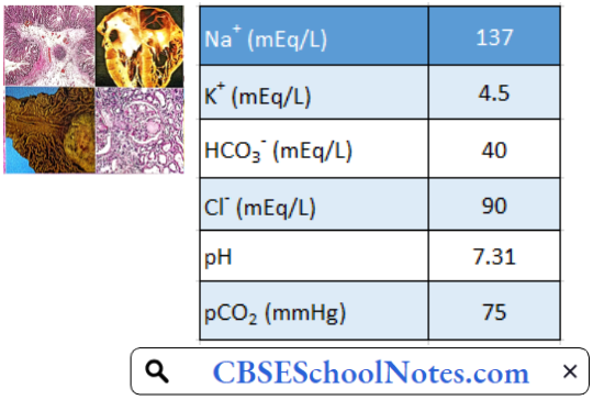
Hypertension Definition: Hypertension is defined as arterial blood pressure greater than 140 mmHg and/or diastolic pressure greater than 90 mmHg. A systolic pressure of 120-140 mmHg or diastolic pressure of 80-90 mmHg is known as prehypertension.

Read and Learn More Pathophysiology

In a vast majority of cases of hypertension, no definite cause can be detected. Such patients are said to suffer from “essential hypertension.” In a small percentage of patients with hypertension, a definite cause can be found, such as kidney disease or/and endocrine disorder. Such cases are said to suffer from secondary hypertension.

Hypertension Symptoms: In most of the cases of high blood pressure, there are no symptoms till the complications occur. When blood pressure is very high, severe headaches may be reported.

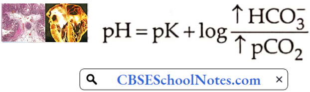
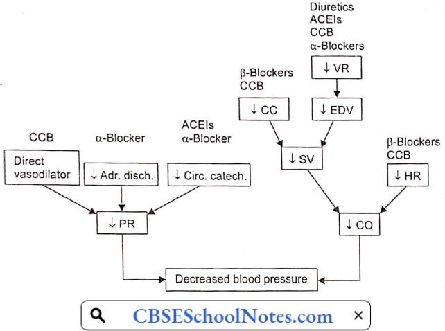
Aetiology And Pathogenesis Of Essential Hypertension: Blood pressure = Cardiac output x Peripheral resistance = CO x PR

Regardless of the origin of hypertension, the actual increase in arterial blood pressure is caused by either an increase in peripheral vascular resistance (PR) or an increase in cardiac output (CO). The former is determined by the vascular tone (i.e. state of constriction) of systemic resistance vessels, whereas the latter is determined by heart rate and stroke volume.

In later stages of hypertension, only peripheral resistance is found to be increased; CO is normal. However, as discussed below, in the early stages of hypertension, many individuals have increased sympathetic activity leading to increased CO.

  1. Genetic Predisposition: Epidemiological studies have shown the importance of genetic predisposition in the development of essential hypertension. If a family history of hypertension is present, the subject has a 3-4 fold greater chance of developing hypertension, at an age earlier than the general population.
    • Although genetics appears to contribute to essential hypertension, the exact mechanism has not been established. Genetic factors interact with environmental factors such as high salt intake, male sex, smoking, obesity, stress and physical inactivity, etc.
  2. Sympathetic Overactivity: There is evidence for a widespread autonomic abnormality in the early phases of hypertension. Overwhelming and excessive sympathetic activity is consistently present in some patients since their childhood.
    • In the early stages of hypertension, blood pressure is elevated when recorded in a doctor’s clinic, but found to be normal when recorded at home. Such cases are said to suffer from “white coat hypertension”. Earlier, it was believed that persons with “white coat hypertension” do not develop established hypertension. There is no support for such an assertion; in fact, such patients are at a high risk of future accelerated hypertension.
    • The hallmark of sympathetic over-activity in these patients is the so-called hyperkinetic state that is best characterized by borderline elevation of blood pressure, a fast heart rate, and an increased cardiac output even at rest.
    • Both the hyperkinetic state and sympathetic overactivity are less readily recognizable later in the course of hypertension. A large proportion of previously hyperkinetic patients later develop established hypertension.
    • It is not clear how from a fast heart rate /high cardiac output form of borderline hypertension is transformed later into the normal cardiac output/high vascular resistance profile that is characteristic of established hypertension.
  3. Role Of Sodium Intake: Essential hypertension is seen primarily in societies with an average sodium intake above 100 mEq/ day (2.3 g sodium); it is rare in societies with average sodium intakes of less than 50 mEq/day (1.2 g sodium). These epidemiological observations led to the suggestion that the development of hypertension requires a threshold level of sodium intake. This factor effect appears to be independent of other risk factors for hypertension, such as obesity.
  4. Vascular Hyper-reactivity: Hypertensive patients manifest greater vasoconstrictive response to infused norepinephrine or immersion of one hand in ice-cold water (cold pressor test) than normal individuals.
    • Greater vasoconstrictive response to norepinephrine has also been demonstrated in normotensive offspring of hypertensive patients as compared to controls with no family history of hypertension. It suggests that vascular hyperreactivity may be genetic in origin.
  5. Renin-Angiotensin-Aldosterone-System (RAAS): In patients with essential hypertension, about 15% have mildly elevated plasma renin activity. In another 60% of hypertensives, plasma renin activity is “within the normal range’ but it may be inappropriate in the presence of elevated blood pressure.
    • Less than 25% patients of with essential hypertension have subnormal plasma renin activity. Moreover, favorable therapeutic response to RAAS blockers suggests that a renin-dependent mechanism may be involved in tire pathogenesis in about 70% of cases of essential hypertension.
    • The fundamental cause of elevated renin activity in such cases is not yet clear. It could be due to chronic sympathetic overactivity. This possibility is supported by the reports that administration of β-blockers in cases with essential hypertension leads to a decrease in plasma renin activity paralleled by a decrease in arterial blood pressure.
  6. Endothelial dysfunction: Due to its position between the bloodstream and vascular smooth muscle, endothelial dysfunction could either be a consequence or a causative factor in essential hypertension. In recent years, considerable evidence has suggested that changes in vascular endothelial function may cause an increase in vascular tone.
    • For example, in hypertensive patients, the vascular endothelium produces less nitric oxide (intrinsic vasodilator). Moreover, the vascular smooth muscle is less sensitive to the actions of this powerful vasodilator. There may also be an increase in endothelin (a vasoconstrictor) production by the endothelial cells, which can enhance vasoconstrictor tone.

Complications Of Untreated Essential Hypertension

  1. Atherosclerosis: Many of the complications of hypertension are related to the effects of sustained elevations of blood pressure on vasculature and heart. Atherosclerosis is commonly associated with and is accelerated by long-standing hypertension.
    • Most of the adverse outcomes in hypertension are associated with thrombosis rather than bleeding. Atherosclerosis predisposes the hypertensive patient to coronary thrombosis and cerebral stroke. Cerebral strokes are more often due to thrombosis rather than hemorrhage in the cerebral vessels.
    • The excess morbidity and mortality related to hypertension are progressive over the whole range of systolic and diastolic blood pressures and are not limited to high values only. However, target-organ damage varies markedly between individuals with similar levels of hypertension. Atherosclerosis may also result in aortic aneurysm or peripheral arterial disease.
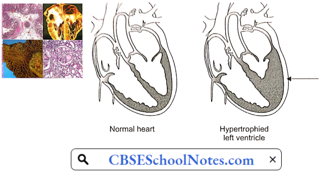
  2. Hypertensive Cardiomyopathy: A sustained increase in blood pressure (afterload) results in hypertrophy and subsequent dilatation of the left ventricle. Electrocardiographic evidence of left ventricular hypertrophy is found in up to 15% of persons with chronic hypertension. Left ventricular hypertrophy may cause or facilitate many cardiac complications of hypertension, including myocardial ischemia, congestive heart failure, and ventricular arrhythmias.
    • Cerebrovascular Complications: Hypertension is an important risk factor for cerebral stroke. Approximately 85% of strokes are due to thrombosis and the remainder are due to hemorrhage in cerebral blood vessels.
    • The term hypertensive encephalopathy is used to describe a group of symptoms and signs that sometimes follow a sudden and sustained rise in blood pressure. The symptoms are characterized by a severe headache, restlessness, impaired judgment, and memory, confusion, somnolence, and stupor. If the condition is not treated, these neurological symptoms may worsen and ultimately turn into a coma.
    • Cerebral encephalopathy seems to result from a failure of autoregulation of cerebral blood flow. The autoregulation seems to fail when hypertension becomes excessive.
  3. Retinopathy: The primary response of the retinal arterioles to systemic hypertension is vasoconstriction. However, sustained hypertension leads to disruption of the blood—retinal barrier, increased vascular permeability, and secondary arteriolosclerosis. Loss of vision may occur.
  4. Renal Complications: Renal failure is one of the important complications of chronic hypertension. Sustained elevation of blood pressure damages renal microvasculature. Renal damage itself is a cause of hypertension (see secondary hypertension below), starting a vicious cycle.
  5. Sexual Dysfunction: Sexual dysfunction is more common and more severe in men with hypertension than it is in the general population. Hypertension is itself the major cause of erectile dysfunction. Experimental studies indicate that essential hypertension results in structural and functional changes in penile vasculature.
    • Cavernous vessels are affected by chronic elevation of arterial blood pressure in the same fashion as other blood vessels. Marked hypertrophy in the smooth muscle of cavernous vessels, increased smooth muscle layer in cavernous space, and increased extracellular matrix (collagen) explain the pathophysiological mechanism of erectile dysfunction in essential hypertension.

Pathophysiological Basis Of Treatment Of Essential Hypertension

Non-Pharmacological Measures

  • Reduction or elimination of factors such as stress, smoking, obesity
  • Regular aerobic exercise
  • Restriction of dietary calories, salt, cholesterol, and saturated fats

Pharmacological Measures: A variety of drugs are being used in the treatment of essential hypertension. They reduce cardiac output, peripheral resistance, or both.

Secondary Hypertension: Secondary hypertension is defined as hypertension that is caused by an underlying well-defined primary cause. It is much less common than essential hypertension, affecting only 5% of hypertensive patients. Some of the causes of secondary hypertension are treatable.

Mechanism Of Action Of Anti Hypertensive Drugs

Secondary Hypertension Symptoms: Like primary hypertension, secondary hypertension usually has no specific signs or symptoms, even if blood pressure has reached dangerously high levels.

Secondary Hypertension Causes

  • Chronic kidney disease (chronic glomerulonephritis)
  • Renal artery stenosis
  • Cushing syndrome (increased secretion of cortisol by a tumor of the adrenal cortex).
  • Aldosteronism (increased secretion of aldosterone by a tumor of the adrenal cortex).
  • Pheochromocytoma (increased secretion of norepinephrine and epinephrine by a tumor of the adrenal medulla).
  • Coarctation of the aorta: There is congenital narrowing (coarctation) in the thoracic aorta. Hypertension is recorded in the upper part of the body only.

Atherosclerosis

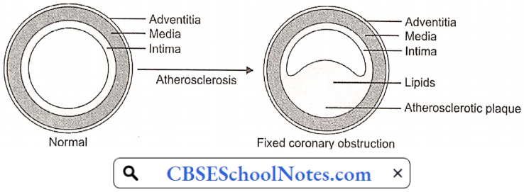
Arteriosclerosis is the thickening, hardening, and loss of elasticity of the walls of arteries. This process gradually restricts the blood flow to one’s organs and tissues and can lead to severe organ damage. Atherosclerosis, which is a specific form of arteriosclerosis, is caused by the build-up of fatty acid-cholesterol plaques in the artery walls.

Atherosclerosis: Atherosclerosis develops primarily in large elastic arteries, for example, the aorta and carotid arteries, or large or medium-sized muscular arteries such as coronary, cerebral, renal, and popliteal arteries.

Atherosclerosis can lead to serious complications such as coronary artery disease, cerebral stroke, and peripheral arterial disease (gangrene of feet or legs). Risk factors associated with atherosclerosis, include:

  • Elevated blood cholesterol and triglyceride levels
  • High blood pressure
  • Obesity
  • Smoking
  • Physical inactivity
  • Diabetes mellitus

Atherosclerosis Symptoms: Atherosclerosis develops gradually. Mild atherosclerosis usually does not have any symptoms. Symptoms develop only when the narrowing of the artery is so severe that an adequate amount of blood does not reach the tissues or organs. At that time symptoms depend on the artery affected.

  • Coronary Artery: Symptoms of angina or myocardial infarction
  • Cerebral Artery: Symptoms of cerebral stroke.
  • Renal Artery: Development of hypertension or symptoms of renal failure.
  • Popliteal Artery: Pain in the legs while walking (claudication)

Pathogenesis Of Atherosclerosis: Aetiological factors named above induce hypercholesterolemia, which disturbs vascular homeostasis, including a decrease in nitrous oxide bioactivity, an increase in superoxide production, an increase in adhesion molecules, and attenuation of endothelium-dependent vasodilatation.

The earliest pathologic lesion of atherosclerosis is the fatty streak. The fatty streak is the result of focal accumulation of serum lipoproteins within the intima of the vessel wall. Gradually, the fatty streak progresses to form a fibrous plaque.

Pathogenesis Of Atheromatous Plaque

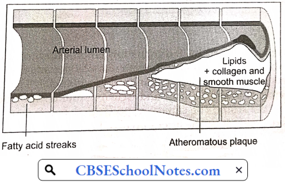
Circulating monocytes infiltrate the intima of the vessel wall. The combination of diabetes and hypertension appears to have an additive effect on monocyte adhesion. These tissue macrophages act as scavenger cells, taking up LDL cholesterol and forming the characteristic foam cells of early atherosclerosis.

These activated macrophages produce numerous factors that are injurious to the endothelium. Atherosclerotic plaque is the result of progressive lipid accumulation along with migration and proliferation of smooth muscle cells. Growth of the fibrous plaque results in progressive luminal narrowing.

Microscopy reveals lipid-laden macrophages, T-lymphocytes, and smooth muscle cells in varying proportions. Atheromatous plaques convert the smooth lining of the tunica intima of the blood vessels to a roughened surface prone to thrombosis.

Aorta Smooth Iner Surface And Atheromatous Inner Surface

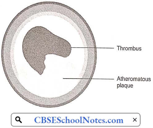
Moreover, developing atherosclerotic plaques are prone to necrosis. Loss of the overlying endothelium or rupture of the protective fibrous cap may result in exposure of the thrombogenic contents of the core of the plaque to the circulating blood.

This exposure constitutes an advanced or complicated lesion. A plaque rupture may result in thrombus formation leading to partial or complete occlusion of the blood vessel.

Complications Of Atherosclerosis

Thrombosis: Rupture of plaque is followed by thrombus formation (intravascular clotting). The rough endothelial lining of a blood vessel attracts platelet adhesion and activation. Activated platelets result in the formation of an intravascular clot.

The thrombus results in critical narrowing of the arterial lumen and ischemia (deficient blood supply) in the tissues supplied by the artery. The clinical response to ischemia caused by obstructive atherosclerosis is dependent on the artery involved:

Coronary Artery Disease

Cardiovascular diseases are the number 1 cause of death globally. An estimated 17.9 million people died from CVDs in 2016, representing 31% of all global deaths. Of these deaths, 85% are due to heart attack and stroke.

In India, studies have reported an increasing prevalence of coronary artery disease over the last 60 years, from 1% to 9-10% in urban populations and <1% to 4-6% in rural populations.

Myocardial Oxygen Supply

  1. The myocardial oxygen supply depends on:
  2. The oxygen content of the arterial blood, and

The rate of coronary blood flow. The oxygen content of arterial blood may be decreased because of decreased hemoglobin concentration or because of poor systemic blood oxygenation (hypoxic hypoxia). Thus, angina may be a presenting feature of a patient with severe anemia or lung disease.

  • In the absence of anemia or lung disease, oxygen supply to the heart is determined by the rate of coronary blood flow.
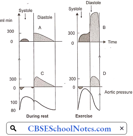
  • In most other organs, because of the greater pressure head, blood flow is greater during systole than in diastole of the heart. However, in the case of the myocardium, the reverse is true. The coronary arteries that run on the surface of the heart are called epicardial coronary arteries.
  • Branches of epicardial arteries that run into and supply blood to the myocardium are called subendocardial coronary vessels. During systole, myocardial contraction has a strangulating effect on the blood vessels passing through the cardiac muscle fibers. Because of this, blood flow in the subendocardial vessels stops.
  • As a result, most myocardial perfusion occurs during diastole when the subendocardial coronary vessels are patent because of the absence of extramural pressure.
  • Although coronary vessels are supplied with sympathetic and parasympathetic nerve fibers, coronary vascular resistance is chiefly determined by intrinsic metabolic factors rather than neural control.
  • Local vasodilator metabolites such as adenosine (chiefly) and other products of anoxic metabolism (lactate, H+, certain prostaglandins) regulate coronary blood flow by a direct action on vascular smooth muscle. During exercise, greater release of local vasodilator metabolites assures greater blood flow in the coronary arteries.
  • Atherosclerotic narrowing of coronary arteries produces its effects mainly by hypo-perfusion (decreased blood supply) of the myocardium. The effect may range from angina to myocardial infarction.

Myocardial Oxygen Supply

Angina Pectoris: In normal individuals during exercise, by the local metabolite control, coronary arteriolar resistance decreases in proportion to the increase in O2 demand of the myocardium. Thus, coronary blood flow increases in proportion to the oxygen demand of the myocardium.

  • When atherosclerotic narrowing is greater than 60-70%, coronary blood flow cannot increase during exercise in spite of the presence of vasodilator metabolites. Therefore, myocardial ischemia results; which is commonly intermittent (only during exertion).
  • Anginal pain is characterized by the fact that it occurs only at times of increased myocardial oxygen demand such as exertion or emotional excitement, but subsides by rest. Such a condition is known as angina. In angina, pain may be localized to the substernum or referred to the left arm, neck, or jaw.

Critical Narrowing Of Coronary Artery Causing Angina

Angina Pectoris Symptoms: Angina symptoms include

  • Pain or discomfort can spread to the chest, jaw, shoulders, arms (mostly the left arm), and back.
  • Chest tightness, burning, heaviness, feeling of squeezing or not being able to breathe.
  • Angina will sometimes cause dizziness, paleness, and weakness.

The Mechanisms Of Cardiac Pain: It is presumed that pain of angina pectoris results from the release of anoxic metabolites (adenosine, bradykinin) by the myocardium. These metabolites excite the sensory ends of the sympathetic and vagal afferent fibers supplying the heart.

Within the spinal cord, cardiac sympathetic afferent impulses may converge with impulses from somatic thoracic structures, which may be the basis for referred cardiac pain, for example, to the left arm.

Location Of Pain In Angina

Pathophysiologic Basis Of Treatment Of Angina: For the immediate relief of angina pain, the patient is advised to take a sublingual tablet of nitroglycerin.

  • Nitroglycerin is converted to a powerful vasodilator nitric oxide (NO) in the body. It may produce some dilation of coronary arteries, but the major action is venous vasodilation. Vasodilation causes the pooling of blood within the venous system, reducing preload to the heart.
  • This causes a decrease in cardiac work, and cardiac oxygen demand and hence relieves angina pain.

Myocardial Infarction

When the myocardial ischemia progresses to a degree that irreversible necrosis of a part of the myocardium occurs, an acute myocardial infarction (MI) is said to have occurred. An acute MI almost always results from an acute thrombotic obstruction of an atherosclerotic coronary artery.

Myocardial Infarction Symptoms

  1. Chest pain or discomfort, possibly described as pressure, squeezing, burning, or fullness
  2. Pain in the left arm, neck, jaw, shoulder, or back accompanying chest pain
  3. Nausea
  4. Fatigue
  5. Shortness of breath
  6. Sweating
  7. Dizziness

In acute MI, the pain has the same characteristics as angina, but it is far more severe, lasts longer, may radiate more widely, and is not relieved by rest or nitroglycerin. Pain may be due to the accumulation of anoxic metabolites as well as products of tissue necrosis. The pain is accompanied by greater psychogenic effects, i.e. feeling of impending death.

Coronary Thrombosis Over Atherosclerotic Plaque

Myocardial Infarction Pathogenesis: Acute myocardial infarction (MI) indicates irreversible myocardial injury resulting in necrosis of a significant portion of myocardium (generally >1 cm). Myocardial infarction is usually due to thrombotic occlusion of a coronary vessel caused by rupture of a vulnerable plaque.

Ischemia induces profound metabolic and ionic perturbations in the affected myocardium. Prolonged myocardial ischemia results in ischemic necrosis of the myocardium. The adult mammalian heart has negligible regenerative capacity, thus the infarcted myocardium heals through the formation of a scar.

Myocardial Infraction

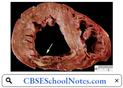
Infarct healing is intertwined with the geometric remodeling of the chamber, characterized by dilation, hypertrophy of viable segments, and progressive dysfunction. Cell membrane damage in acute MI leads to the release of certain intracellular enzymes. An increase in their plasma levels is used as diagnostic evidence of myocardial infarction.

Histology Of Myocardial Infract

Myocardial Infarction Risk Factors

  • Age: Men age 45 or older and women age 55 or older are more likely to have a heart attack than are younger men and women.
  • Tobacco: This includes smoking and long-term exposure to second-hand smoke.
  • High Blood Pressure: Over time, high blood pressure can damage coronary arteries.
  • High Blood Cholesterol Or Triglyceride Levels: A high level of low-density lipoprotein (LDL) cholesterol (the ‘bad’ cholesterol) is most likely to narrow arteries. A high level of triglycerides, a type of blood fat related to your diet, also increases your risk of heart attack.
  • Obesity: Obesity is associated with high blood cholesterol levels, high triglyceride levels, high blood pressure, and diabetes.
  • Diabetes.
  • Family history of heart attack
  • Lack of physical activity
  • Stress.

Myocardial Infarction Diagnosis

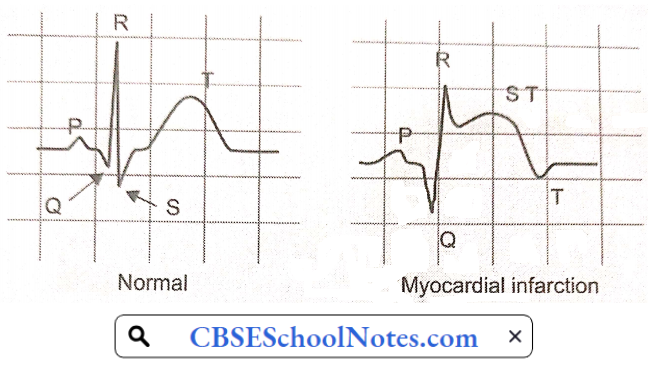
  1. Electrocardiogram: An electrocardiogram (ECG) is a recording of the electrical activity of the heart. Abnormalities in electrical activity usually occur with heart attacks and can identify the areas of the heart muscle that are deprived of oxygen and/or areas of muscle that have died.
    • In a patient with typical symptoms of heart attack (such as crushing chest pain) and characteristic changes of heart attack on the ECG, a secure diagnosis of heart attack can be made quickly in the emergency room and treatment can be started immediately.
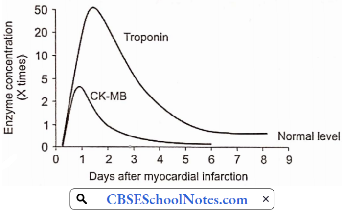
  2. Blood Tests: Cardiac enzymes are proteins that are released into the blood by dying heart muscles. These cardiac enzymes are creatine kinase (CK-MB), and troponin, and their levels can be measured in blood.
    • These cardiac enzymes typically are elevated in the blood several hours after the onset of a heart attack. Currently, troponin levels are considered the preferred lab tests to use to help diagnose a heart attack, as they are indicators of cardiac muscle injury or death.

A series of blood tests for tire enzymes performed over a 24-hour period are useful not only in confirming the diagnosis of heart attack, but the changes in their levels over time also correlate with the amount of heart muscle that has died.

ECG In Myocardial Infraction

Serum Enzyme Levels In Myocardial Infarction

Complications Of MI: More important complications include

  • Cardiogenic shock
  • Cardiac arrhythmias (ventricular tachycardia or ventricular fibrillation are life-threatening complications).
  • Congestive heart failure

Pathophysiologic Basis Of Treatment: Treatment aims at immediate restoration of blood flow in the artery blocked by a blood clot followed by measures to prevent thrombosis again

  • Angioplasty And Stent: Special tubing with an attached deflated balloon is threaded up to the coronary arteries. A stent is a wire mesh tube used to prop open an artery during angioplasty.
  • Long-term antiplatelet therapy

Congestive Heart Failure

Congestive heart failure (CHF), or heart failure is defined as an inability of the heart to pump blood at a rate appropriate for the metabolic requirements of the tissues;

  • Aetiology: Important causes include:
  • Hypertension: Due to high blood pressure, expulsion of blood requires more forceful ventricular contraction during each systole. Over time, the left ventricle initially undergoes hypertrophy and later it dilates leading to heart failure.
  • Coronary Artery Disease: Myocardial infarction weakens the myocardium.
  • Valvular Disease: Disorders of aortic or mitral valve cause extra burden on the heart. Initially, the heart undergoes hypertrophy and later dilatation.

Congestive Heart Failure Symptoms And Signs

  1. Exercise intolerance
  2. Fatigue
  3. Dyspnoea on effort
  4. Cyanosis
  5. Ankle edema
  6. Distended neck veins
  7. Liver enlargement (hepatomegaly)
  8. Spleen enlargement (splenomegaly)
  9. Oedema feet
  10. Ascites (fluid in the peritoneal cavity, abdomen)

Signs Of Right Sides Heart Failure

Pathophysiology Of CHF: When the heart is unable to pump out a sufficient amount of blood, a number of natural compensatory mechanisms are activated so as to improve the cardiac output and maintain normal perfusion of the vital organs. These mechanisms include:

  • Frank-Starling mechanism
  • Increased adrenergic discharge
  • Regional redistribution of cardiac output
  • Hormonal mechanisms

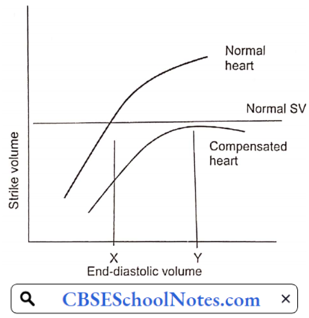
As you have learned in physiology lectures, cardiac output can be increased by an increase in end-diastolic volume (EDV) (Starling mechanism) as well as an increase in sympathetic discharge to the heart. Both of these mechanisms are utilized to bring the failing heart to produce normal cardiac output.

The other two compensatory mechanisms help in these primary mechanisms are:

  1. Frank-Starling Mechanism: By working at greater EDV (Y), the heart can pump out a normal amount of blood. EDV is increased by the fourth compensatory mechanism mentioned above.
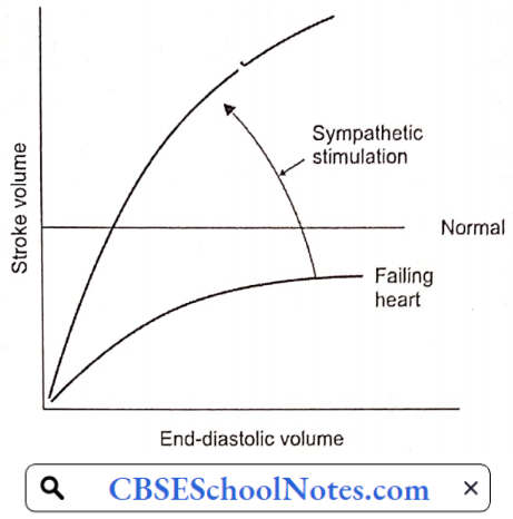
  2. Increased Adrenergic Discharge: In the failing heart, depressed cardiac output is sensed by high-pressure baroreceptors located in the carotid sinus and aortic arch, leading to a reflex increase in adrenergic discharge to the heart and blood vessels.
    • In the heart, increased adrenergic discharge improves the cardiac output by increasing the heart rate as well as stroke volume. In the blood vessels, it causes a redistribution of cardiac output described next.
  3. Redistribution Of Cardiac Output: The redistribution of cardiac output serves as an important compensatory mechanism when cardiac output is reduced. Blood flow is redistributed so that the delivery of oxygen to vital organs, such as the brain and myocardium, is maintained at normal or near-normal levels, while flow to less critical areas, such as the cutaneous and muscular beds and viscera, is reduced. Vasoconstriction mediated by the adrenergic nervous system is largely responsible for this redistribution.
  4. Hormonal Mechanisms: Decreased cardiac output activates the renin-angiotensin II mechanism. Angiotensin II produces arteriolar constriction and increases thirst (increased water intake). It also increases aldosterone secretion, thereby producing salt and water retention in the kidney. As a result of both these factors, blood volume and thereby EDV is increased.

Mechanisms Of Compensation Of A failing Heart By Frank Starling Mechanism

Effect Of Increased Synpathetic Discharge On Stroke Volume In A Failing Heart

Congestive Heart Failure Decompensation: As the cardiac function gradually deteriorates, the compensatory mechanisms discussed above cannot maintain normal cardiac output. Moreover, excessive increase in EDV increases left and right atrial pressures resulting in congestion in the lungs and in systemic veins. At this point, congestive heart failure is said to have set in.

Pathophysiological Basis of Treatment

  1. Strengthening The Force Of Contraction Of The Heart: Digitalis.
  2. Reducing Salt And Water Retention: Diuretics
  3. Reducing Sympathetic Over-Activity: Beta-blockers

Cell injury and Cellular Adaptation Notes

Basic Principles Of Cell Injury And Adaptation

Cell Injury And Adaptation Definitions: Aetiology is the origin of a disease, including the underlying causes and modifying factors.

  • Pathogenesis refers to the steps in the development of disease. It describes how aetiologic factors trigger cellular and molecular changes that give rise to the specific functional and structural abnormalities of the disease. Whereas etiology refers to why a disease arises, pathogenesis describes how a disease develops.
  • Pathology describes the structural changes observed in a diseased tissue or an organ.
  • Pathophysiology describes the biochemical, and functional changes that occur in cells, tissues, and organs in response to injury.
  • Morphology describes changes in the gross (naked eye) or microscopic appearance of a tissue or an organ.

Read and Learn More Pathophysiology

Cell Injury And Adaptation Symptoms And Signs: A symptom is a subjective feeling of a departure from normal function that is apparent to a patient, reflecting the presence of an unusual state, or of a disease. A symptom can be subjective (felt by the patient) or objective (seen by the patient).

Tiredness is a subjective symptom, whereas cough or fever are objective symptoms. In contrast to a symptom, a sign is a clue to a disease elicited by an examiner or a doctor. For example, edema feet is a sign of congestive heart failure or liver failure.

Homeostasis Physiology

The mammalian cells are very sensitive. The cell’s survival and function is dependent on the maintenance of the internal environment. The composition of the internal environment may be disturbed by a variety of external or internal factors. Examples of external factors that may disturb the internal environment include exposure to extremes of heat or cold, absence of food, deficiency of oxygen, etc.

  • Examples of internal factors that may disturb the internal environment include infections, blood loss, increased utilization of glucose, and production of excess lactic acid by vigorously contracting muscles during severe exercise, etc.
  • In nil such conditions, various organs of the body maintain homeostasis, that is, act in a harmonious fashion and prevent marked changes in the physical and chemical composition of extracellular fluid. Failure of the homeostatic mechanism results in disturbed body function known as disease.

Homeostatic Mechanisms: Various physiological or pathological processes tend to disturb one or more components of the internal environment. The disturbance sets into motion a series of physiological responses that eliminate the disturbing factor and normalize the composition of extracellular fluid. The homeostatic mechanisms allow an organism to function effectively in a broad range of environmental conditions.

Basic Principles Of Cell Injury And Adaptation Basic Homeostatic Mechanism Of Homeostasis

All homeostatic mechanisms have at least three components:

  1. Receptor that detects a change in the internal environment and sends information to the control center.
  2. The control center is the structure that evaluates the disturbance and activates the correcting mechanisms.
  3. The effector is the structure that carries out the corrective responses as directed by the control center.

For example, exposure to cold tends to lower the body temperature. The change in body temperature is detected by cold thermal receptors (receptors). The information is communicated to the temperature-regulating areas of the hypothalamus in the brain (center). The body responds by involuntary contractions of skeletal muscle called shivering (effector).

  • Shivering generates heat and prevents a fall in body temperature. Exposure to hot environment tends to raise body temperature. The change in body temperature is detected by warmth thermal receptors (receptors).
  • The information is communicated to the temperature-regulating areas of the hypothalamus in the brain (center). The body responds by sweating which results in heat loss by evaporation (effector). This is an example of body temperature homeostasis.

Basic Principles Of Cell Injury And Adaptation Components Of Homeostatic Mechanisms

Homeostasis Physiological Control Systems

Our body consists of many organ systems, and each organ system consists of more than one organ. Therefore, central control mechanisms are required so as to allow coordination of activity of different organs of an organ system and coordination between different organ systems. Our body has two major control systems.

  1. Neural Control: It is chiefly exerted on skeletal muscles and exocrine glands. The response occurs in milliseconds.
  2. Endocrine Control: It is chiefly exerted on metabolic reactions. The response may take seconds, hours, or even days.

Open-Loop And Closed-Loop Processes (Systems): Physiological processes may be governed by an open-loop system or a closed-loop system. In an open-loop system, the output product (signal) has no control over the process.

The entire process is controlled by the input signals. In a closed-loop control, the product in the system (output signal) has an effect on the input signal. This is called a feedback phenomenon. Feedback control may be a negative feedback or a positive feedback system.

1. Open-Loop System: The presence of food in the intestine results in the secretion of enzymes from the pancreas. however, the concentration of pancreatic enzymes in the intestine has no effect on the secretion of pancreatic acini. This is an example of an open-loop system.

Basic Principles Of Cell Injury And Adaptation Types Of Physiological Systems

2. Negative Feedback Control: In the negative feedback control system, the output signal has an inhibitory effect on the input signal. For example, ingestion of food results in the secretion of gastric juice that contains hydrochloric acid (secreted by parietal cells of gastric glands).

  • Secretion of acid has a negative feedback effect on parietal cells—when the concentration of acid in gastric juice reaches a certain level (pH 2), the acidity has an inhibitory effect on parietal cells and thus further acid secretion stops.
  • As a result, gastric acidity cannot increase beyond a certain predetermined degree. Negative feedback control mechanisms are most often used in the maintenance of homeostasis. This system prevents deviations from a given set point.

Basic Principles Of Cell Injury And Adaptation A Physiological Closed Loop Negative Feedback System

3. Positive Feedback System: In a positive feedback system, the output signal accentuates the input signal. As a result, the output deviates more and more from the initial output. The positive feedback system is not involved in homeostasis. It is used to accelerate or reinforce a reaction.

  • For example, during delivery, at about 40 weeks of pregnancy, uterine contractions start that are weak to begin with. Mild uterine contractions cause pressure of the head of the baby on the cervix and dilate it. Dilatation of the cervix reflexly causes the release of the hormone oxytocin by the pituitary gland.
  • Oxytocin causes stronger uterine contractions that cause further dilation of the cervix and still greater of oxytocin. In this way, by a positive feedback mechanism, uterine contractions gradually become so strong that the baby is expelled out of the uterus.

Basic Principles Of Cell Injury And Adaptation A Positive Feedback System During Delivery

4. Feedforward System: This system allows the human body to foresee a change in the environment and prepares the body for the change. The information is sent ahead of time to the control system. The effector system is modified before any change has taken place, i.e. by anticipating change in the environment.

Basic Principles Of Cell Injury And Adaptation Feedforward Control System

A Feedforward control system is advantageous in fast reactions. For example, during a cricket match, when a fielder tries to catch the ball, he has to anticipate the trajectory of the ball by visual input and run to an appropriate place. In most of the voluntary movements, both feedforward and negative feedback systems operate simultaneously.

Cellular Response To Pathological Stress Or Noxious Stimuli

Cells or tissues encounter pathological stresses or stimuli, in the form of hypoxia, ischemia, infections, and physical or chemical insults. The cells can respond to such stimuli by undergoing two fundamental processes:

  1. Adaptation, modifying themselves to a new steady state and preserving viability and function. The principal adaptive responses are hypertrophy, hyperplasia, atrophy, and metaplasia.
  2. Cell Injury: If the stressful stimulus exceeds the adaptive capability, cell injury results. Depending on the strength of noxious stimulus, cell injury may be
    • Reversible And Subsequently Normal Cellular Function Is Restored, Or
    • Irreversible, Resulting In Death Of The Cells Or Tissues

Basic Principles Of Cell Injury And Adaptation Cellular Responses To Stressful Or Injurious Stimuli

Cellular Adaptations To Stress

Adaptations are reversible changes in the number, size, or metabolic activity of cells in response to changes in their environment.

  • Physiologic adaptations usually represent responses of cells to normal stimulation by hormones or endogenous chemical mediators (for example, the hormone-induced enlargement of the breast and uterus during pregnancy).
  • Pathologic adaptations are responses to stress that allow cells to modulate their structure and function and thus escape injury. Such adaptations can take several distinct forms, namely hypertrophy, hyperplasia, atrophy, and metaplasia.

Basic Principles Of Cell Injury And Adaptation Various Types Of Pathologic Adaptations In Response To Stress

Hypertrophy Heart : Hypertrophy is an increase in the size of cells resulting in increase in the size of the organ. In pure hypertrophy, there are no new cells, just bigger cells containing increased amounts of structural proteins and organelles. Hypertrophy occurs when cells have a limited capacity to divide.

Basic Principles Of Cell Injury And Adaptation Pathologic Hypertrophy In The Heart

Hypertrophy can be physiologic or pathologic and is caused either by increased functional demand or by growth factor or hormonal stimulation. An example of physiologic hypertrophy is the enlarged muscle of the weightlifter. Thickening of ventricular muscle in response to the narrowing of the outlet valve is an example of pathologic hypertrophy.

Hyperplasia Uterus : Hyperplasia is characterized by an increase in the size of an organ due to an increase in cell number. It results from a proliferation of tissue cells. Hyperplasia takes place if the tissue contains cell populations capable of replication; it may occur concurrently with hypertrophy and often in response to the same stimuli. Hyperplasia can be physiologic or pathologic.

  • An example of physiologic hyperplasia is the enlargement of the mammary gland by the proliferation of the glandular tissue during pregnancy and lactation. Benign prostatic hyperplasia leading to enlargement of the prostate gland is an example of pathological hyperplasia.
  • An important point is that in all of these situations, the hyperplastic process remains controlled; if the signals that initiate it disappear, the hyperplasia disappears. It is this responsiveness to normal regulatory control mechanisms that distinguishes pathologic hyperplasia from cancer, in which the growth control mechanisms become dysregulated or ineffective.
  • Hypertrophy and hyperplasia can occur simultaneously. The massive physiologic enlargement of the uterus during pregnancy occurs as a consequence of estrogen-stimulated smooth muscle hypertrophy and smooth muscle hyperplasia.

Basic Principles Of Cell Injury And Adaptation Hyperplasia Of Mammary Gland During Pregnancy And Lactation

Atrophy: Shrinkage in the size of the cell by the loss of cell substance is known as atrophy. When a sufficient number of cells are involved, the entire tissue or organ diminishes in size, becoming atrophic.

  • Although atrophic cells may have diminished function, they are not dead. Causes of atrophy include a decreased workload (for example immobilization of a limb to permit healing of a fracture), loss of innervation, diminished blood supply, inadequate nutrition, loss of endocrine stimulation, and aging (senile atrophy).
  • Although some of these stimuli are physiologic (for example the loss of hormone stimulation in menopause) and others pathologic (for example denervation). Cellular atrophy represents a retreat by the cell to a smaller size at which survival is still possible; a new equilibrium is achieved between cell size and diminished blood supply or nutrition.

Basic Principles Of Cell Injury And Adaptation Atrophy Of The Muscles Of The Palm Of Hand

Metaplasia: Metaplasia is a reversible change in which one cell type (epithelial or mesenchymal) is replaced by another cell type. In this type of cellular adaptation, a cell type sensitive to a particular stress is replaced by another cell type that is better able to withstand the adverse environment.

  • Epithelial metaplasia is exemplified by the change of normal stratified squamous epithelium of the lower end of the esophagus to the intestinal columnar epithelium in patients of gastro-oesophageal reflux disease (GERD).
  • In GERD, weak functioning of the lower oesophageal sphincter leads to the reflux of gastric acid into the esophagus. Chronic acid exposure converts stratified squamous epithelium to columnar epithelium. The columnar epithelium is more resistant to acid.
  • Another example is the metaplasia seen in the respiratory mucosa in chronic smokers. In this case the normal delicate ciliated columnar epithelial cells of the trachea and bronchi a tv locally replaced by more rugged stratified squamous epithelial cells. however. the influences that induce metaplastic change, if persistent, may predispose to malignant transformation of the epithelium.

Basic Principles Of Cell Injury And Adaptation Metaplasis In Oesophageal Epithelium

Cell Injury

Cell injury results when cells are stressed so severely that they are no longer able to adapt or when cells are exposed to inherently damaging agents or suffer from intrinsic abnormalities (for example in DNA or proteins). Different injurious stimuli affect many metabolic pathways and cellular organelles. Injury may progress through a reversible stage and culminate in irreversible change, i.e. cell death.

Reversible Cell Injury: In early stages or mild forms of injury, the functional and morphologic changes are reversible if the damaging stimulus is removed. At this stage, although there may be significant structural and functional abnormalities, the injury has typically not progressed to severe membrane damage and nuclear dissolution.

Irreversible Cell Injury (Cell Death): Cell death may occur in the form of

  1. Necrosis, or
  2. Apoptosis
  1. Necrosis: With continuing damage, the injury becomes irreversible, at which time the cell cannot recover and it dies. When damage to membranes is severe, enzymes leak out of lysosomes, enter the cytoplasm, and digest the cell, resulting in necrosis. Cellular contents also leak through the damaged plasma membrane into the extracellular space, where they elicit a host reaction (inflammation).
    • Necrosis is the major pathway of cell death in many commonly encountered injuries, such as those resulting from ischemia, exposure to toxins, various infections, and trauma.
  2. Apoptosis: When a cell is deprived of growth factors, or the cell’s DNA or proteins are damaged beyond repair, the cell kills itself by another type of death, called apoptosis, which is characterized by nuclear dissolution without complete loss of membrane integrity.
    • Whereas necrosis is always a pathologic process, apoptosis serves many normal-time ions amt is not necessarily associated with pathologic cell injury.

Basic Principles Of Cell Injury And Adaptation Stages Of Response To Cell Injury

Furthermore, in keeping with its role in certain physiologic processes, apoptosis does not elicit an inflammatory response.

Histologic Signs Of Reversible Cell Injury

  1. Cellular Swelling: The first sign of almost all forms of injury to cells, is a reversible alteration called cellular swelling. Microscopic examination may reveal small, clear vacuoles within the cytoplasm; these represent distended and pinched-off segments of the endoplasmic reticulum (ER). This pattern of nonlethal injury is sometimes called vacuolar degeneration. The intracellular changes associated with reversible injury include:
    • Plasma membrane alterations such as blunting, or distortion of microvilli, and loosening of intercellular attachments;
    • Mitochondrial swelling
    • Dilation of the er with detachment of ribosomes and
    • Minimal nuclear alterations (clumping of chromatin).
    • Cellular swelling is the result of the failure of energy-dependent ion pumps in the plasma membrane, leading to an inability to maintain ionic and fluid homeostasis.
  2. Fatty Change: Fatty change occurs in hypoxic injury and in various forms of toxic or metabolic injury. It is manifested by the appearance of small or large lipid vacuoles in the cytoplasm. It is principally encountered in cells participating in fat metabolism (for example hepatocytes). Like cellular swelling, fatty change is also the reversible stage of tissue injury.

Basic Principles Of Cell Injury And Adaptation Fatty Change In The Liver

Histologic Signs Of Necrosis: Necrosis is the type of cell death that is associated with loss of membrane integrity and leakage of cellular contents culminating in the dissolution of cells, largely resulting from the degradative action of enzymes on lethally injured cells.

  • The leaked cellular contents often elicit a local host reaction, called inflammation, that attempts to eliminate the dead cells and start the subsequent repair process.
  • The enzymes responsible for the digestion of the cell may be derived from the lysosomes of the dying cells themselves and from the lysosomes of leukocytes that are recruited as part of the inflammatory reaction to the dead cells

Necrosis is characterized by changes in the cytoplasm and nuclei of the injured cells

  1. Cytoplasmic Changes: Increased eosinophilia (deep pink staining in slides stained with H and E). It is attributable in part to the increased binding of eosin to denatured cytoplasmic proteins and in part to the loss of the basophilia that is normally imparted by the ribonucleic acid (RNA) in the cytoplasm.
    • Compared with viable cells, the cell may have a more glassy, homogeneous appearance, mostly because of the loss of glycogen particles.
    • When enzymes have digested cytoplasmic organelles, the cytoplasm becomes vacuolated and appears “moth-eaten.”
    • By electron microscopy, necrotic cells are characterized by discontinuities in plasma and organelle membranes, marked dilation of mitochondria with the appearance of large amorphous densities, and disruption of lysosomes.
  2. Nuclear Changes: Nuclear changes assume one of three patterns, all due to the breakdown of DNA and chromatin.
    • Karyolysis, in which basophilia of the chromatin may fade presumably secondary to deoxyribonuclease (DNase) activity.
    • Pyknosis is characterized by nuclear shrinkage and increased basophilia; the DNA condenses into a solid shrunken mass.
    • Karyorrhexis, the pyknotic nucleus undergoes fragmentation.

Basic Principles Of Cell Injury And Adaptation Nuclear Changes In Necrosis

Fates Of Necrotic Cells

  • Necrotic cells may persist for some time or maybe digested by enzymes and disappear.
  • Dead cells may be phagocytosed by other cells
  • Dead cells ultimately become calcified.

Mechanisms Of Cell Injury: Cell injury results from functional and biochemical abnormalities in one or more of several essential cellular components. The principal targets and biochemical mechanisms of cell injury are

  1. Mitochondria and their ability to generate ATP and ROS under pathologic conditions
  2. Disturbance in calcium homeostasis
  3. Damage to cellular (plasma and lysosomal) membranes
  4. Damage to DNA.
  1. ATP Depletion: The major causes of ATP depletion are reduced supply of oxygen and nutrients, as well as mitochondrial damage. ATP is required for virtually all synthetic and degradative processes within the cell, including membrane transport, protein synthesis, lipogenesis, etc. Deficiency of ATP leads to
    • Reduced activity of Na-K-ATPase pump resulting in intracellular accumulation of sodium and efflux of potassium. The net gain of solute is accompanied by the gain of water, causing cell swelling and dilation of the endoplasmic reticulum.
    • A compensatory increase in anaerobic glycolysis in an attempt to maintain the cell’s energy sources. As a consequence, intracellular glycogen stores are rapidly depleted, and lactic acid accumulates, leading to decreased intracellular pH and decreased activity of many cellular enzymes.
  2. Influx Of Calcium: Ischaemia and certain toxins cause an increase in cytosolic calcium concentration, initially because of the release of Ca2+ from the intracellular stores, and later resulting from the increased influx of Ca2+ across the plasma membrane due to failure of ATP-dependent Ca2+ pumps. Increased cytosolic Ca2+ activates a number of enzymes, with potentially deleterious cellular effects.
    • There is structural disruption of the protein synthetic apparatus, manifested as the detachment of ribosomes from the rough endoplasmic reticulum. Thus, there is a reduction in protein synthesis. Ultimately, there is irreversible damage to mitochondrial and lysosomal membranes, and the cell undergoes necrosis.
  3. Defects In Membrane Permeability: Increased membrane permeability leading ultimately to overt membrane damage is a consistent feature of most forms of cell injury that culminate in necrosis. The most important sites of membrane damage during cell injury are the mitochondrial membrane, the plasma membrane, and the membranes of lysosomes.
    • Mitochondrial membrane damage. Damage to mitochondrial membranes leads to a further reduction in the production of ATP culminating in necrosis.
    • Plasma membrane damage. Plasma membrane damage leads to loss of osmotic balance and influx of fluids and ions, as well as loss of cellular contents.
    • Injury to lysosomal membranes results in leakage of their enzymes into the cytoplasm and activation of the acid hydrolases in the acidic intracellular pH of the injured (for example ischaemic) cell. Lysosomes contain ribonucleases (RNAses), DNAses, proteases, glucosidases, and other enzymes. Activation of these enzymes leads to enzymatic digestion of cell components, and the cells die by necrosis.
  4. Damage To DNA: Cells have mechanisms that repair damage to DNA, but if this damage is too severe to be corrected, the cell initiates its suicide program and dies by apoptosis.

Apoptosis Pathway

Apoptosis is a pathway of cell death in which cells activate enzymes that degrade the cells’ own nuclear DNA and nuclear and cytoplasmic proteins. Fragments of the apoptotic cells then break off, giving the appearance that is responsible for the name (apoptosis, “falling off”). The plasma membrane of the apoptotic cell remains intact, but the membrane is altered in such a way that the cell and its fragments become targets for phagocytes.

Basic Principles Of Cell Injury And Adaptation Necrosis And Apoptosis

The dead cell and its fragments are rapidly cleared before cellular contents have leaked out, so apoptotic cell death does not elicit an inflammatory reaction in the host. Apoptosis differs in this respect from necrosis, which is characterized by loss of membrane integrity, enzymatic digestion of cells, leakage of cellular contents, and frequently a host reaction. However, apoptosis and necrosis sometimes coexist, and apoptosis induced by some pathologic stimuli may progress to necrosis.

Causes Of Apoptosis: Apoptosis occurs in many normal situations and serves to eliminate potentially harmful cells and cells that have outlived their usefulness. It also occurs as a pathologic event when cells are damaged beyond repair, especially when the damage affects the cell’s DNA; in these situations, the irreparably damaged cell is eliminated by apoptosis.

Physiological Apoptosis

  1. Involution of hormone-dependent tissues upon hormone deprivation, such as endometrial cell breakdown during the menstrual cycle, and regression of the lactating breast after weaning.
  2. Cell loss in proliferating cell populations, such as intestinal crypt epithelia, in order to maintain a constant number.
  3. Elimination of cells that have served their useful purpose, such as neutrophils in an acute inflammatory response.

Pathologic Apoptosis

  1. Damaged DNA
  2. Apoptosis eliminates cells that are genetically altered or injured beyond repair and does so without eliciting a severe host reaction, thereby keeping the extent of tissue damage to a minimum. Death by apoptosis is responsible for the loss of cells in a variety of pathologic states that damage the DNA of cells.
  3. Cell injury in certain infections, particularly viral infections, in which loss of infected cells is largely due to apoptotic death that may be induced by the virus.

Intracellular Accumulations

Intracellular accumulations of various substances and extracellular deposition of calcium, both of which are often associated with cell injury.

Fat: fatty change refers to any abnormal accumulation of triglycerides within parenchymal cells. It is most often seen in the liver since this is the major organ involved in fat metabolism, but it may also occur in the heart, skeletal muscle, kidney, and other organs.

Steatosis may be caused by toxins, protein malnutrition, diabetes mellitus, obesity, or anoxia. Alcohol abuse and diabetes associated with obesity are the most common causes of fatty change in the liver (fatty liver).

Cholesterol And Cholesteryl Esters: Cellular cholesterol metabolism is tightly regulated to ensure normal cell membrane synthesis without significant intracellular accumulation. However; phagocytic cells may become overloaded with lipids (triglycerides, cholesterol, and cholesteryl esters) in several different pathologic processes. Of these, atherosclerosis is the most important.

Glycogen: Excessive intracellular deposits of glycogen are associated with abnormalities in the metabolism of either glucose or glycogen. In poorly controlled diabetes mellitus, the prime example of abnormal glucose metabolism, is glycogen accumulates in renal tubular epithelium, cardiac myocytes, and p cells of the islets of Langerhans.

Basic Principles Of Cell Injury And Adaptation Glycogen Deposition In The Hepatocytes In A Case Of Diabetes Mellitus

Lipofuscin: It is an insoluble brownish-yellow granular intracellular material that accumulates in a variety of tissues (particularly the heart, liver, and brain) as a result of age or atrophy.

Basic Principles Of Cell Injury And Adaptation Lipofuscin Granules In A Neuron

Calcium: Pathologic calcification is a common process in a wide variety of disease states; it implies the abnormal deposition of calcium salts. When the deposition occurs in dead or dying tissues, it is called dystrophic calcification; it occurs in the absence of derangements in calcium metabolism, i.e. with normal serum levels of calcium. In contrast, the deposition of calcium salts in normal tissues is known as metastatic calcification and is almost always secondary to hypercalcemia.

Basic Principles Of Cell Injury And Adaptation Dystrophic And Metastatic Calcification

Acidosis And Alkalosis

The Concept Of pH: An acid is a chemical species that can donate a proton (H+), and a base is a species that can accept (gain) a proton. Pure water undergoes an extremely small degree of dissociation to yield H+ and OH.

H2O ⇔ H+ + OH

The concentration of H+ in water is 10-7 mEq/L. Water is regarded as neutral. Acids are solutions with H+ concentration greater than 10-7 (for example 10-6 or 10-5 mEq/L). Alkalis or bases are solutions with H+ concentration less than 10-7 (for example 10-8 or 10-9 mEq/L). Expression of H+ concentration in the body fluids as described above is cumbersome; hence a symbol pH came to be used:

Basic Principles Of Cell Injury And Adaptation The Concept Of pH

  • Thus pH of pure water is written as 7. The arterial blood has an average pH of 7.4 (normal range 7.35-7.45). A decrease in arterial pH value below 7.35 is known as acidosis, whereas the term alkalosis is used to describe arterial pH values higher than 7.45.
  • Arterial pH values below 6.8 or above 8 are not compatible with life. In fact, arterial pH is maintained within a narrow range, transiently with the help of acid-base buffers in the body fluids, and finally by the kidneys and the lungs.

Regulation Of Hydrogen Ion Balance

  1. Buffer systems response—very rapid (in seconds), incomplete
  2. Respiratory responses—rapid (in minutes), incomplete
  3. Renal responses—slow (in hours to days), complete

Under normal circumstances, tremendous amounts of hydrogen ions (H+) are continuously added to the body fluids. Carbon dioxide accounts for the addition of over 12,000 mEq H+ per day. Nonvolatile (fixed) acids account for another 60 mEq/ day.

  • Almost all the CO2 is excreted by the lungs, whereas the kidney is responsible for the excretion of non-volatile acid products of protein metabolism. Lactic acid produced during severe exercise or keto acids produced in severe diabetes is also excreted by the kidney.
  • Fruits are the main dietary source of the alkali. They contain sodium and potassium salts of weak organic acids, whose metabolism produces NaHCO3 or KHCO3 (and CO2). Normally, the alkali content of the diet is very small and all the normal individuals excrete acidic urine except for transient post-prandial alkaline tide.

Role Of Respiration: In response to changes in blood pH, respiratory responses occur within minutes by stimulation/depression of respiratory centers in the CNS.

In spite of the addition of 12,000 mEq H+ per day to the blood, the pH of arterial blood remains remarkably constant at 7.4. Similarly, pCO2 of the arterial blood is kept constant at 40 mm Hg. This is made possible by two factors:

  1. In the venous blood, CO2 is converted to H2CO3 and further to H+ and HCO3. Hydrogen ions are immediately buffered by the blood buffers, chiefly hemoglobin and plasma proteins.
  2. As the venous blood passes through the lungs, CO2 is regenerated by reversal of the reactions mentioned above 1 and excreted very efficiently. Pulmonary ventilation is so delicately adjusted that it exactly matches the CO2 produced in the body.
    • Even during severe exercise, when CO2 production increases 20-fold, CO2 excretion is so efficient that arterial pCO2 does not increase at all. Such a delicate control is made possible by the fact that ventilation is controlled by both CO2 as well as H+ concentration through central and peripheral chemoreceptors.
    • Effect Of CO2: CO2 is a highly diffusible gas. It can easily cross the blood-brain and blood-CSF barriers, and stimulate the medullary central chemoreceptors. In contrast, H+ cannot cross these barriers easily. Therefore, central chemoreceptors are most sensitive to changes in arterial pCO2 and less so to changes in H+ concentration.
    • Effect Of pH: An increase in H+ concentration of arterial blood also stimulates pulmonary ventilation, chiefly through the peripheral (sino-aortic) chemoreceptors. Therefore, the respiratory system helps in the regulation of the acid-base balance of the body even when the increase in H+ concentration is not due to CO2 but due to non-volatile acids like sulphuric acid, phosphoric acid, or lactic acid.

Role Of Kidneys: The kidneys regulate pH by either acidification or alkalinization of the urine. The renal response occurs over hours/days and is capable of nearly complete restoration of acid-base balance. As mentioned above, about 60 mEq of H+ is added to the blood every day as nonvolatile acids.

  • They cannot be excreted by the lungs. They are excreted by the kidneys in an indirect manner. In the kidneys, most of the excretory products are initially filtered into the glomerular filtrate. All that is necessary for their excretion is that renal tubules reabsorb them partially or not all.
  • In contrast, the concentration of H+ in the blood is so small, that they cannot be excreted in this manner. Actually, most of the H+ produced in the body does not remain as such. They are immediately buffered by HCO3 and other buffers. The kidney generates new hydrogen ions equivalent to the amount metabolically produced and actively secretes them into the urinary tubules, where they are buffered by phosphate and ammonium ions.
  • The generation of H+ in the renal tubules is accompanied by the production of HCO3 which diffuses into the blood circulation and replenishes the amount of HCO3 lost during the initial buffering of the acids. In case an excess of base (NaHCO3) is ingested, it is excreted by the kidney by filtration followed by partial or complete non-reabsorption.
  • In primary pulmonary diseases such as emphysema, pulmonary excretion of CO2 is diminished, and therefore arterial pCO2 and H+ concentrations tend to rise (respiratory acidosis). In such circumstances, renal excretion of H+ is the only means of maintaining body pH near normal (renal compensation).
  • Similarly in chronic renal failure, renal excretion of H+ is diminished leading to metabolic acidosis. In such a condition, excessive loss of CO2 by hyperventilation is the only means of maintaining body pH near normal (respiratory compensation).

Anion Gap Concept: In the plasma, total cations (Na+, K+, Ca++, Mg++, etc.) are always counterbalanced by total anions (Cl, HCO3, PO4, SO4, etc). Of these ions, only Na+, K+, Cl, and HCO3 are routinely measured. Therefore, the concentration of measured anions is always less than the concentration of measured cations. The difference is known as the anion gap

Anion gap = {[Na3] + [K3]} – {[Cl] + [HCO3] )

Anion Gap Example: Anion gap = {140 mEq/L + 4 mEq/L } – {100 mEq/L + 28 mEq/L} = 16 mEq/L

  • The anion gap concept is useful in the differential diagnosis of metabolic acidosis. In one group of disorders producing metabolic acidosis, the anion gap becomes larger than normal. Such disorders are said to produce high anion gap metabolic acidosis.
  • In the other group of disorders, the anion gap remains normal. Such disorders are said to produce normal attire gap metabolic acidosis. Estimation of the anion gap is also a useful tool to assess mixed acid-base disorders.

In clinical practice, since K+ concentration does not vary grossly, the anion gap is usually calculated as follows:

Anion gap = [Na+] – { [Cl] + [HCO3,] }

Calculated in this way, the normal anion gap is 12 ± 4 mEq/L.

Pathophysiology Of Acid Base Disorders

1. Metabolic Acidosis (Ma): The primary abnormality in metabolic acidosis is a decline in plasma HCO3 concentration. This metabolic abnormality may arise because of:

  1. Increased H+ load on the body in the form of lactic acidosis, ketoacidosis, or ammonium chloride administration.
  2. Deficient renal H+ excretion.
  3. Loss of HCO3from GI tract or kidneys.

Metabolic Acidosis May Be Classified Into Two Major Groups:

  1. High anion gap MA and
  2. Normal anion gap (hyperchloraemic) MA.

High Anion Gap MA: There are three prominent causes of high anion gap MA:

  1. Lactacidosis
    • Circulatory shock
    • Severe hypoxia
  2. Ketoacidosis
    • Diabetic ketoacidosis
    • Alcoholic ketoacidosis
    • Starvation
  3. Renal failure
    • Acute or chronic

In the uncompensated stage of such MA, the acid-base picture is:

Basic Principles Of Cell Injury And Adaptation Metabolic Acidosis Formula

Acidosis stimulates central and peripheral chemoreceptors causing hyperventilation (respiratory compensation). The renal compensation consists of the excretion of highly acidic urine (↑H excretion). Renal compensation is not possible if MA is due to renal failure.

The respiratory compensation decreases the arterial pCO2, whereas renal compensation generates HCO3, (a side effect of H+ secretion), which partially restores the depleted HCO3. The acid-base status of a case of compensated MA is as follows:

Basic Principles Of Cell Injury And Adaptation Reneal Compenstation

The two most important causes of high anion gap MA in clinical practice are diabetic ketoacidosis and renal failure. The two disorders can be differentiated by the study of serum K+ level, which is elevated in renal failure and subnormal in diabetic ketoacidosis. A typical pattern of acid-base status and electrolyte status of patients of these two types of disorders is given below:

Basic Principles Of Cell Injury And Adaptation Pattern Of Acid Base Status And Electrolyte

Normal Anion Gap (Hyperchloremic) MA

  1. Diarrhea
  2. Renal tubular acidosis

High anion gap MA is characterized by a decrease in plasma HCO3 level, but no significant change in plasma Cl level. In the hyperchloraemic type of MA, a decrease in plasma HCO3 is accompanied by a significant increase in plasma Cl hence the name hyperchloraemic.

This type of MA is typically seen in patients suffering from diarrhea or renal tubular acidosis (RTA). In diarrhea, HCO3 is lost from the gut in exchange for Cl. In RTA, the failure of bicarbonate reabsorption from the renal tubules results in greater reabsorption of Cl. In either case, the plasma Cl level is significantly elevated. A typical pattern of acid-base status and electrolyte pattern in the case of RTA is given below

Basic Principles Of Cell Injury And Adaptation Normal Anion Gap

The anion gap in the three cases with MA given above is calculated below:

Anion gap = [Na+] – [(Cl) 4-(HCO3)]

Diabetic: 125 – [90 + 5] = 30 mEq/L (high anion gap)

Uremic: 135 – [101 + 12] = 22 mEq/L (high anion gap)

RTA: 140 – 1115 + 15] = 10 mEq/L (normal anion gap)

2. Metabolic Alkalosis

  1. Excessive sodium bicarbonate ingestion
  2. Persistent vomiting

Metabolic alkalosis occurs as a result of net gain of bicarbonate (for example ingestion of NaHCO3 for peptic ulcer) or more often due to loss of non-volatile acids (for example HCl in prolonged vomiting). The primary acid-base picture in the uncompensated stage is as follows:

Basic Principles Of Cell Injury And Adaptation Metabolic Alkalosis

For metabolic alkalosis, there is respiratory as well as renal compensation. Alkalosis inhibits the peripheral chemoreceptors, resulting in hypoventilation and elevation of pCO2 In the kidney, metabolic alkalosis results in decreased secretion of H+ by the renal tubules. Hence filtered bicarbonate is not completely absorbed.

The urinary losses of bicarbonate decrease the extent of elevation of plasma bicarbonate. Hence the change in pH of blood is minimized. The acid-base status of a case of compensated metabolic alkalosis is as follows:

Basic Principles Of Cell Injury And Adaptation Rebal Compensation Results In Elevation

Metabolic alkalosis due to persistent vomiting is accompanied by not only the loss of acid but also fluid from the stomach. The resulting hypovolemia becomes a strong stimulus for sodium reabsorption in the kidneys through Na+ -K+ as well as Na+-H antiport systems.

Therefore, when metabolic alkalosis is accompanied by hypovolemia, the renal response tends to aggravate alkalosis rather than correct it. It leads to hypokalemia as well. A fairly typical pattern of acid-base status and electrolytes in a patient with persistent vomiting is given below:

Basic Principles Of Cell Injury And Adaptation Fairly Pattern Of Acid Base Status And Electrolytes Patient

3. Respiratory Acidosis: The primary abnormality in respiratory acidosis is the elevation of PaCO2 due to alveolar hypoventilation. Alveolar hypoventilation also reduces PaO2. Therefore, hypoxemia always accompanies hypercapnia.

Basic Principles Of Cell Injury And Adaptation Respiratory Acidosis

Acute respiratory acidosis is defined as hypercapnia developed in the time prior to renal compensation, i.e. less than 24 hours. In acute respiratory acidosis, the acid-base status is as follows:

Acute Respiratory Acidosis May Be Caused By:

  • Acute airway obstruction (severe asthma)
  • Central respiratory drive depression
    • Dings: Narcotics, benzodiazepines, barbiturates
    • Neurologic Disorders: Encephalitis, brainstem disease, trauma, poliomyelitis.

Chronic respiratory acidosis is most commonly present in

  • Chronic obstructive pulmonary disease
    • Emphysema
    • Chronic bronchitis
  • Chest wall deformities
    • Kyphoscoliosis

The renal compensation involves ↑H+ excretion, as well as increased generation of HCO3 The resultant increase in plasma bicarbonate concentration partially restores the blood pH towards normal:

Basic Principles Of Cell Injury And Adaptation Respiratory Acidosis

Renal compensation results in the elevation of plasma HCO3by 3.5 mEq/L for every 10 mmHg increase in pCO2 A fairly typical pattern of acid-base and electrolyte status of a patient with chronic respiratory acidosis is given below:

Basic Principles Of Cell Injury And Adaptation Renal Compensation Resulsts In Elevation

4. Respiratory Alkalosis: Respiratory alkalosis is the most common acid-base disorder in a critically ill patient. It is primarily caused by alveolar hyperventilation leading to decreased paCO2.

Basic Principles Of Cell Injury And Adaptation Respiratory Alkalosis

Hypoxia due to acute pulmonary disease (for example pneumonia) or chronic pulmonary disease, sepsis, and psychogenic hyperventilation are common causes of respiratory alkalosis.

Renal compensation to decreased arterial pCO2 is decreased renal secretion of H+.

Basic Principles Of Cell Injury And Adaptation Reneal Compenstation

Consequently, bicarbonate is not generated in the kidneys. More importantly, even the filtered bicarbonate is not fully reabsorbed. Hence plasma HCO3 falls markedly which minimizes the change in blood pH:

A fairly typical pattern of acid-base and electrolytes in a patient with respiratory alkalosis is given below

Basic Principles Of Cell Injury And Adaptation Acid Base And Electrolytes In A patient With Respiratory Alkalosis

Electrolyte Imbalance

Electrolyte Imbalance is an abnormality in the concentration of electrolytes in the body. Electrolytes play a vital role in maintaining homeostasis within the body. They help to regulate heart and neurological function, fluid balance, oxygen delivery, acid-base balance and much more.

Electrolyte imbalances can develop by consuming too little or too much electrolyte as well as excreting too little or too much electrolyte. The most serious electrolyte disturbances involve abnormalities in the levels of sodium, potassium or calcium.

1. Sodium

  • Hypernatraemia: Hypernatraemia means that the concentration of sodium in the blood is too high. An individual is considered to have high sodium at levels above 145 mEq/L of sodium.
    • Hypernatraemia Causes
      • Inadequate water consumption
      • Severe dehydration
      • Excessive loss of bodily fluids as a result of prolonged vomiting, diarrhea, sweating, or respiratory illness
      • Certain medications, such as diuretics and corticosteroids
    • Hypernatraemia Symptoms
      • Dehydration
      • Nausea
      • Vomiting
      • Fatigue
      • Weakness
      • Increases thirst
  • Hyponatremia: Hyponatremia is defined as a concentration lower than 135 mEq/L.
    • Hyponatremia Causes
      • Excessive fluid loss through the skin from sweating or burns
      • Vomiting or diarrhoea
      • Overhydration
      • Congestive heart, or kidney failure
      • Certain medications, including diuretics
      • Syndrome of inappropriate secretion of antidiuretic hormone (SIADH)
    • Hyponatremia Symptom: The severity of symptoms is directly correlated with the severity of hyponatremia and rapidness of onset.
      • Loss of appetite
      • Agitation
      • Nausea
      • Weakness
      • Vomiting
      • Seizures, coma, and death
      • Confusion

2. Potassium

  • Hyperkalaemia: Hyperkalaemia means the concentration of potassium in the blood is >5 mEq/L.
    • Hyperkalemia Causes:
      • Kidney failure
      • Severe acidosis, including diabetic ketoacidosis
      • Certain medications, including some blood pressure medications and diuretics
      • Adrenal insufficiency
    • Hyperkalaemia Symptoms
      • Nausea
      • Vomiting
      • Diarrhea
      • Muscle cramps
      • Numbness
      • Tingling
      • Absence of reflexes
      • Paralysis
      • Cardiac arrhythmias can result in death.
  • Hypokalaemia: Hypokalaemia is defined as the plasma concentration of potassium is <3.5 mEq/L.
    • Hypokalaemia Causes
      • Severe vomiting or diarrhea
      • Dehydration
      • Certain medications, including laxatives, diuretics, and corticosteroids
    • Hypokalaemia Symptoms
      • Muscle weakness
      • Cramping
      • Cardiac arrhythmias

3. Calcium

  • Hypercalcaemia: Hypercalcaemia is when plasma calcium concentration is above 10.5 mEq/dL.
    • Hypercalcemia Causes
      • Hyperparathyroidism
      • Malignancy
      • Hyperthyroidism
    • Hypercalcaemia Symptoms
      • Abdominal pain
      • Constipation
      • Kidney stones
  • Hypocalcaemia: Hypocalcaemia is defined as a plasma calcium level less than 9 mg/dL ‘
    • Hypercalcemia Causes
      • Vitamin D deficiency
      • Hypoparathyroidism
      • Multiple blood transfusions
    • Hypercalcaemia Symptoms
      • Muscle cramping or twitching
      • Numbness around the mouth and fingers
      • Arrhythmias

Acute And Chronic Inflammation Notes

Basic Mechanism Involved In The Process Of Inflammation And Repair

Inflammation: Inflammation is the complex biological response of vascular tissues to harmful stimuli, such as pathogens, damaged cells, or irritants. Avascular tissues such as cornea, articular cartilage, intervertebral disc, etc. do not show inflammatory response.

  • The inflammatory response is a protective attempt by the organism to remove the injurious stimuli as well as initiate the healing process. Inflammation is not a synonym for infection. Even in cases where inflammation is caused by infection, the two are not synonymous: Infection is caused by an exogenous pathogen, while inflammation is the response of the organism to the pathogen.
  • Inflammation can be classified as either acute or chronic. Acute inflammation is the initial response of the body to harmful stimuli and is achieved by the increased movement of plasma and leukocytes (initially neutrophils) from the blood into the injured tissues.
  • A cascade of biochemical events propagates and matures the inflammatory response, involving the local vascular system, the immune system, and various cells within the injured tissue. It comes to an end within a few hours or days.
  • Prolonged inflammation persisting for weeks or months is known as chronic inflammation. Whereas, neutrophil accumulation in the lesion is a hallmark of acute inflammation, chronic inflammatory lesion is characterized by the presence of lymphocytes, monocytes, macrophages, and plasma cells.

Read and Learn More Pathophysiology

Another hallmark of chronic inflammation is the simultaneous processes of tissue destruction and healing resulting in the formation of scar tissue. Inflammation may result from two sets of causes: Exogenous and endogenous:

  • Inflammation Exogenous Factors
    • Mechanical injury (traumatic injury),
    • Physical injury (extremely low or high temperature, ionizing irradiation, microwaves)
    • Chemical injury (caustic agents, poisons, venoms, etc.)
    • Biological injury (viruses, microorganisms, protozoan and metazoan parasites).
    • Ischaemic injury
  • Inflammation Endogenous Factors
    • The immunopathological responses such as allergic inflammations and autoimmune inflammatory disorders
    • Endogenous products of tissue metabolism such as gout.

Acute Inflammation

Acute Inflammation Cardinal Signs: Acute inflammation is a short-term process/ usually appearing in a few minutes or hours and ceasing once the injurious stimulus has been removed. It is characterized by five cardinal signs

  • Redness
  • Warmth
  • Swelling
  • Pain
  • Loss of function

The first four (classical signs) were described by Celsus about 2000 years ago, while loss of function was added to the list later by Virchow in 1870.

  • Redness and warmth are due to increased blood flow at body core temperature to the areas such as skin, which normally are at a lower temperature; swelling is caused by the accumulation of fluid and plasma proteins in the extravascular spaces; pain is due to the release of chemicals that stimulate pain nerve endings or sensitize them to other stimuli.
  • Loss of function has multiple causes, chiefly pain, and local edema.
  • These five signs appear when acute inflammation occurs on the body’s surface. In the case of acute inflammation of internal organs, all five signs may not be apparent.

Acute Inflammatory Response: The acute inflammatory response may be discussed under two headings:

  1. The vascular response and
  2. The cellular response.

1. The Vascular Response: Alterations in the microvasculature (arterioles, capillaries, and venules) of the injured tissue are the earliest response to the injury. It consists of hemodynamic changes and changes in vascular permeability.

  • Hemodynamic Changes: Transient vasoconstriction of the arterioles and reduced blood flow is the immediate response irrespective of the type of injury. It usually lasts only a few seconds but may be prolonged up to five minutes if the injury is very severe. It is followed by:
    • Persistent and progressive vasodilatation begins in the arterioles and spreads to the capillaries and venules as well. This change becomes prominent within an hour of injury. Vasodilatation results in increased blood flow to the microvasculature and accounts for tire clinical signs of redness and warmth.
      • Vasodilatation is brought about by the release of vasodilator mediators by the injured tissue cells as well as by the blood cells attracted by the injury.
    • Transudation of fluid into the extracellular space (edema) is another consequence of vasodilatation. Starling forces, chiefly capillary hydrostatic pressure and plasma protein oncotic pressure govern the tissue fluid exchange across the capillary wall.
      • It involves tissue fluid formation at the proximal segment of the capillary followed by reabsorption in the distal segment. Vasodilatation, by increasing the capillary hydrostatic pressure shifts the balance of the Starling forces in favor of greater exudation and decreased reabsorption. Thus, local edema results.
    • Stasis. Loss of fluid from the capillaries leads to increased viscosity of blood flowing through the capillaries, with resultant stasis due to the increase in the concentration of the cells within blood. Stasis allows leukocytes to marginate along die endothelium, a process critical to their recruitment into the tissues.
      • Normal flowing blood prevents this, as the shearing force along the periphery of die vessels moves blood cells into the middle of the vessel. Nutritional supply to die issue may be so compromised that it may become ischemic, even necrotic.
  • Increased Vascular Permeability: All the blood vessels are lined by a continuous layer of endothelial cells, which provide a passive diffusion barrier. It permits the free diffusion of water and solutes but restricts the movement of larger molecules such as plasma proteins and cellular components of blood.
    1. The endothelial cells are joined together by tight junctions. In inflammatory conditions, the excessive fluid transferred into extracellular space consists not only of usual water and solutes (called transudate) but also contains a high concentration of plasma proteins.
      • Such a tiuid is called an exudate. The exudate is formed because of markedly increased vascular permeability. The causes of increased vascular permeability include the following
    2. Opening of endothelial inter-cellular tight junctions, particularly in the postcapillary venules due to contraction of endothelial cells. It is mediated by the release of histamine, bradykinin, and other chemical mediators of inflammation. This response begins immediately after injury and usually lasts for a short duration (15-30 minutes).
    3. Direct injury to endothelial cells results in necrosis and the appearance of physical gaps at the site of detached endothelial cells. This type of increased permeability lasts for hours or even days.
    4. Endothelial injury is also mediated by leukocytes. Margination followed by leukocyte adhesion may result in the activation of leukocytes. The activated leucocytes release proteolytic enzymes and toxic free radicals which cause endothelial injury and increased vascular leakiness.
    5. The capillaries, newly formed during the process of repair, are excessively leaky.

Mediators Of Increased Vascular Permeability: The primary source of vasoactive mediators of increased permeability during an inflammatory process is derived from injured tissue cells as well as plasma

2. The Cellular Response: Inflammatory response, which lasts more than a few hours, is characterized by the accumulation of white blood cells within the area of injury. In bacterial infections, physical or thermal injury, polymorphonuclear neutrophils are first to arrive (“first line of defense”).

Twenty-four to forty-eight hours later, a large number of macrophages can be seen in the inflamed area. In allergic inflammation, eosinophils and mast cells predominate. In viral infections, lymphocytes are the first to arrive.

Polymorphonuclear Neutrophils And Monocyte-Macrophages: The accumulation of neutrophils and monocyte macrophages at the site of inflammation is due to the presence of locally generated chemical mediators called chemotactic factors.

Vasoactive Mediators Of Increased Vascular Permeability During Inflammation

  1. Chemotaxis: The chemotactic-mediated transmigration of leukocytes involves the initial crossing of several barriers (endothelial basement membrane and matrix), followed by transport in the interstitial fluid to the inflamed area. The process is called chemotaxis.
    • The chemical agents that act as potent chemotactic agents include leukotrienes, platelet factor, components of the complement system (C5a in particular), cytokines (IL-8 in particular), soluble bacterial products, monocyte chemo-attractant protein and eotaxin factor (for eosinophils).
    • There is an increasing concentration gradient of chemotactic agents between an adjacent blood capillary the site of inflammation and leukocyte migration follows the gradient.
  2. Margination: This is the first step towards the transmigration of leukocytes out of the blood capillaries. Normal blood flow is characterized by an axial stream of red cells, leukocytes, and platelets and a peripheral cell—a free layer of plasma close to the vessel wall.
    • Due to the slowing of blood flow and stasis, the central stream of cells widens and blood cells including leukocytes come closer to the vessel wall. This phenomenon is known as margination.
  3. Adhesion: Marginated leukocytes tend to stick briefly to the endothelial cells or roll over them. Injury leads to neutralization of the normal charge on the leukocytes and endothelial cells, resulting in a loose transient adhesion of leukocytes to the endothelial cells.
  4. Transmigration (Diapedesis): During chemotactic response, there is a characteristic change in the morphological orientation of the leukocyte (neutrophil or monocyte). It loses its classical rounded appearance and becomes wedge-shaped. At first, the leading edge passes into the space between two adjacent endothelial cells, damaging the basement membrane, and passes out of the vessel wall.
    • By amoeboid movements, the rear part of the cell contains lysosomal granules, and lastly, the nucleus leaves the blood vessel.
  5. Phagocytosis: Neutrophils and macrophages have an inherent capacity to recognize and engulf foreign particles. Coating of the bacteria by plasma proteins containing IgG and/or complement (opsonization) renders them more liable to phagocytosis.
    • When a neutrophil or a macrophage becomes bound to a bacterium (or a foreign particle), there is a localized contraction of the cell under the point of contact, resulting in the formation of a cup-shaped invagination.
    • Through the pseudopodia thrown out at the margins of the cups, the bacterium, enclosed in a vacuole, is internalized into the phagocyte and called a phagosome. Movement of the phagosome towards the granule-rich areas of the cytoplasm results in the fusion of the phagosome to an adjacent lysosome.
    • Next, the lysosomal granules are discharged into the phagosome.
    • This phenomenon is known as degranulation. The lysosomal membrane is incorporated into the vacuole membrane.
    • The resulting structure is called a phagolysosome. Generally, the release of lysosomal granules is restricted to the phagolysosome.
    • However, when the phagosome formation occurs in a granule-rich area or the phagocyte attempts to engulf too large a particle, lysosomal granules may be discharged into extracellular space causing damage to the host tissue cells in the vicinity.
  6. Bacterial Killing And Digestion: This is the ultimate objective of phagocytosis. Anti-microbial agents act by the following two mechanisms
    • Oxygen-Dependent Bactericidal Mechanisms: Degranulation is accompanied by the activation of two enzymes present in the leukocyte granules, namely NADPH oxidase and myeloperoxidase.
      • Activation of NADPH-oxidase is associated with a sharp increase in oxygen consumption in the leukocyte (the respiratory burst) leading to the generation of highly toxic superoxide (O2) and hydrogen peroxide (H2O2). Myeloperoxidase catalyzes the formation of highly toxic hypochlorous acid (HCIO).
    • Oxygen-Independent Bactericidal Mechanisms: Lysosomal granules contain a number of agents that do not require oxygen for their bactericidal activity. These agents include lysosomal hydrolases, permeability-increasing factors, defensins, lysozyme, and cationic protein.

Chemotaxis

Steps In Transport Of A Neutrophil Out Of A Blood Capillary

The Process Of Phagocytosis

Mast Cells/Basophils: Mast cells and basophils play a central role in inflammatory and immediate allergic reactions. They are able to release potent inflammatory mediators, such as histamine, proteases, chemotactic factors, cytokines, and metabolites of arachidonic acid that act on the blood capillaries, smooth muscle, connective tissue, mucous glands, and inflammatory cells.

Both mast cells and basophils contain special cytoplasmic granules which store mediators of inflammation. The extracellular release of the mediators from the mast cells (degranulation) may be induced by:

  1. Physical destruction, such as high temperature, mechanical trauma, ionizing irradiation, etc.;
  2. Chemical substances, such as toxins, venoms, proteases;
  3. Endogenous mediators, including tissue proteases, cationic proteins derived from eosinophils and neutrophils;
  4. Immune mechanisms which may be IgE-dependent or IgE-independent

The increase in the number of mast cells and basophils, and the enhanced secretion at sites of inflammation, can accelerate the elimination of the cause of tissue injury or, paradoxically, may lead to a chronic inflammatory response. Thus, manipulating mast cell and basophil adhesion may be an important strategy for controlling the outcome of allergic and inflammatory responses.

Eosinophils: Eosinophils is a leukocyte that resides predominantly in submucosal tissue and is recruited to sites of specific immune reactions, including allergic diseases. The large specific granules contain four distinct cationic proteins which exert a range of biological effects on host cells and microbial targets: Major basic protein (MBP), eosinophil cationic protein (ECP), eosinophil-derived neurotoxin (EDN), and eosinophil peroxidase (EPO).

  • In addition, histaminase and a variety of hydrolytic lysosomal enzymes are also present in the large specific granules. These proteins have major effects not only on the potential role of eosinophils in host defense against helminthic parasites but also in contributing to tissue dysfunction and damage in eosinophil-related inflammatory and allergic diseases.
  • Compared to neutrophils, eosinophils have limited phagocytic activity which is mainly aimed at killing multicellular parasites. Another possible beneficial function of eosinophils is the inactivation of mediators of anaphylaxis.

Systemic Effects Of Acute Inflammation: Fever is due to the release of interleukin 1 (a cytokine), prostaglandins, or tumor necrosis factor from the inflammatory tissues, either of which can disturb tire hypothalamic temperature regulating center. Fever may also be induced by certain constituents in the cell wall of dead bacteria called pyrogens.

  1. Leucocytosis is a feature of infections or even non-infectious inflammations. Typically, the total leukocyte count is between 15,000 and 20,000 IpL. Usually, in bacterial infections, leucocytosis is due to neutrophilia; in viral infections due to lymphocytosis; and in allergic conditions due to eosinophilia. Some infections, e.g. typhoid fever, however, are associated with leucopenia (neutropenia with relative lymphocytosis).
  2. Lymphangitis: lymphadenitis in the lymph vessels and lymph nodes draining the area of inflammation is commonly seen. These responses represent either a non-specific reaction to chemical mediators released by the inflamed tissues or an immunological response to foreign antigens.
  3. Acute phase proteins: Acute inflammation is commonly accompanied by increased concentrations of several plasma proteins such as C-reactive protein, alpha-2 macroglobulin, and fibrinogen (collectively called acute phase proteins).
    • The precise function of these proteins in inflammation is largely unclear. However, when measured in the laboratory, they can serve as useful markers of inflammation.
    • These proteins also increase the erythrocyte sedimentation rate (ESR), a nonspecific indicator of inflammation. Finally, prolonged or widespread inflammation can deplete complement leading to decreased levels of certain components of complement in the serum.
  4. Other symptoms such as decreased appetite, lactacidosis, negative nitrogen balance, and increased slow-wave sleep are commonly seen in acute infections. Most of these seem to be produced by interleukin 1.
  5. Shock may develop in severe acute inflammatory conditions. Tumor necrosis factor (TNF-α), a cytokine, is one of the mediators of acute inflammation. Bacteraemia/septicemia may result in the release of a massive amount of TNF-α leading to widespread vasodilatation and increased vascular permeability.
    • These changes lead to intravascular volume loss, hypotension, and circulatory shock. Microthrombi may be formed throughout the body which may lead to disseminated intravascular coagulation (DIC), bleeding, and death.

Outcomes Of Acute Inflammation: The acute inflammatory response may have one of the following four outcomes depending on whether or not injury results in significant tissue loss or the inflammatory stimulus is rapidly removed:

  1. Resolution
  2. Healing,
  3. Suppuration, or
  4. Chronic inflammation.
  1. Resolution: Such an outcome follows the complete removal of the agent or microorganism that triggered the inflammatory response. The process includes the removal of any injured (necrotic) host cells. This is the ideal outcome for acute inflammation. It is more likely if cellular damage has been minimal, for example, the resolution of lobar pneumonia.
  2. Healing May Involve Two Processes:
    • Regeneration: The replacement of damaged or lost tissue by normal tissue of a similar type. It occurs only in tissues that contain cells capable of dividing (for example, epithelial tissues such as the epidermis of the skin).
    • Repair: Scar formation, fibrosis. It involves the replacement of damaged or lost tissue by collagen fibers (scar tissue). This is the healing mechanism for those tissues that cannot regenerate (dermis, nerve, muscle, etc.).
  3. Suppuration And Abscess Formation: If there has been a large amount of cellular necrosis, or if there is a great deal of bacterial contamination, exudates and dead leukocytes (pus) can accumulate forming an abscess. In time, connective tissue walls of the abscess and limits its spread. Resolution and healing cannot take place until adequate drainage of the abscess has been provided.
  4. Chronic Inflammation: Normally, the acute inflammatory response to cellular injury has subsided by the time tissue healing begins. If tissue destruction is prolonged, inflammation and attempts at healing occur at the same time. This produces a picture of chronic inflammation.

Mechanism Of Final Resolution Of Acute Inflammation: Acute inflammation normally resolves by mechanisms that have remained somewhat elusive. Emerging evidence now suggests that an active, coordinated program of resolution is initiated in the first few hours after an inflammatory response begins. After entering tissues, granulocytes promote the switch of arachidonic acid—derived prostaglandins and leukotrienes to lipoxins, which initiate the termination sequence.

Neutrophil recruitment thus ceases and programmed death by apoptosis is engaged. Consequently, apoptotic neutrophils undergo phagocytosis by macrophages, leading to neutrophil clearance and release of anti-inflammatory and reparative cytokines such as transforming growth factor-β1. The anti-inflammatory program ends with the departure of macrophages through the lymphatics.

Allergic Inflammation: An allergic reaction is the result of an inappropriate immune response triggering inflammation. A common example is hay fever, which is caused by a hypersensitive response by skin mast cells to allergens.

Pre-sensitized mast cells respond by degranulation, releasing vasoactive chemicals such as histamine. These chemicals propagate an excessive inflammatory response characterized by blood vessel dilation, production of pro-inflammatory molecules, cytokine release, and recruitment of leukocytes. A severe inflammatory response may mature into a systemic response known as anaphylaxis.

Chronic Inflammation

Chronic inflammation occurs when the damaging stimulus persists and the process of continuing tissue necrosis, organization, and repair all occur concurrently. In addition to acute inflammation, the specific defenses of the immune system are activated around the area of damage, and tissues are infiltrated by activated lymphoid cells.

The Chronic Inflammatory Tissue Shows:

  1. Necrotic cell debris
  2. Acute inflammatory exudate
  3. Vascular and fibrous granulation tissue
  4. Lymphoid cells
  5. Macrophages
  6. Collagenous scar

Chronic inflammation may be caused by one of the following three mechanisms:

  1. Chronic Inflammation Following An Acute Inflammation: When tissue destruction is extensive or bacteria survive and persist at the site of inflammation, for example, osteomyelitis or pneumonia leading to a lung abscess.
  2. Recurrent Attacks Of Acute Inflammation: Repeated attacks of acute inflammation may culminate in chronicity of the disease process, for example, pyelonephritis resulting from recurrent attacks of urinary tract infection, or repeated attacks of acute cholecystitis culminating in chronic cholecystitis.
  3. Chronic Inflammation Starting De Novo: In such cases, the inflammatory agent produces a chronic inflammatory response to begin with.

General Features Of Chronic Inflammation: Ordinarily, agents that produce an acute inflammatory response are removed by the neutrophils and macrophages by phagocytosis and digestion. However, certain agents cannot be removed by an acute inflammatory response, for example, Mycobacterium tuberculosis, fungus, or a suture.

The mechanism of dealing with such indigestible agents is termed chronic or granulomatous inflammation. Chronic inflammation response primarily serves to contain the pathological process, as well as to remove the offending agent, if possible.

Though chronic inflammatory responses may somewhat differ in detail, depending on the offending agent, the following features are common to all chronic inflammations:

  1. Mononuclear Cell Infiltration
  2. Tissue Destruction And necrosis
  3. Proliferative Changes.
  1. Mononuclear Cell Infiltration
    1. Macrophages: The macrophages comprise the most important cells in chronic inflammation. These cells are recruited by chemotactic migration from the circulation as well as by local proliferation.
      • Activated macrophages release several biologically active substances such as neutral and acid proteases, oxygen-derived reactive metabolites, and cytokines. These agents bring about tissue destruction, neo-vascularization, and fibrosis. Chronic inflammatory lesions usually show some other chronic inflammatory cells:
    2. Lymphocytes: These cells are a prominent feature of chronic inflammatory lesions. They perform vital functions both in cell-mediated and humoral immune responses. The T-lymphocytes function not only as cytotoxic killer cells but also regulate macrophage recruitment and activation through the secretion of lymphokines (cytokines) and modulate antibody production.
    3. Plasma Cells: These cells are also usually present in a chronic inflammatory lesion. Plasma cells are immune-activated B-lymphocytes rich in cytoplasmic reticulum. These cells are the primary source of antibodies specific to the antigen present at the site of chronic lesions.
  2. Tissue Destruction: It is one of the important features of most of the chronic inflammatory responses. As mentioned above, it is brought about by several biologically active substances released by activated macrophages.
  3. Proliferative Changes: As a result of necrosis, proliferation of small blood vessels is stimulated. Eventually, collagen is laid down and healing by fibrosis occurs.

Histological Appearance Of Acute And Chronic Inflammation

Summary Of Differences Between Acute And Chronic Inflammation

Differences Between Acute And Chronic Inflammation

Basic Principles Of Wound Healing

If there is an injury to the skin, under normal circumstances, the wound heals in a few days. The process of wound healing consists of four overlapping phases:

  1. Hemostasis Phase: Injury to the skin or other tissues results in bleeding. The first step in wound healing involves the stoppage of bleeding (hemostasis). Hemostasis starts when blood leaks out of the body. The circulating platelets collect at the site of injury, stick together, form a platelet plug, and seal the break in the wall of the blood vessel. This step is known as primary hemostasis.
    • However, the platelet is soft and temporary. In the next step known as secondary hemostasis, the blood collected at the site of injury clots. Clotting of blood seals the leak in blood vessels permanently.
    • The blood clot reinforces the platelet plug with threads of fibrin which are like a molecular binding agent. The hemostasis stage of wound healing happens within a few minutes.
  2. Inflammatory Phase: Inflammation is the second stage of wound healing and begins right after the injury when the injured blood vessels lose blood. Inflammation both controls bleeding and prevents infection. During the inflammatory phase, damaged cells are removed from the wound area. Swelling, heat, pain, and redness commonly seen during this stage of wound healing are signs of acute inflammation.
    • If some bacteria get into the wound, blood macrophages remove the pathogens. Inflammation is a natural part of the wound healing process and is only problematic if prolonged or excessive if infection cannot be controlled.
  3. Proliferative Phase: The proliferative phase features three distinct stages:
    • Filling the wound
    • Contraction of the wound margins
    • Covering the wound by epithelialization
    • The gap in the skin is filled by newly built tissue consisting of collagen fibers and extracellular matrix. In addition, a new network of blood vessels grows into the wound bed forming shiny, deep red granulation tissue. When tissues are injured, fibroblasts around the injured region differentiate into myofibroblasts, a type of highly contractile cells that produce abundant extracellular matrix proteins.
    • It has become clear that both fibroblasts and myofibroblasts play a critical role in the wound healing process. Fibroblast cells lay down collagen fibers. Myofibroblasts cause the wound to contract by gripping the wound edges and pulling them together.
    • In healthy stages of wound healing, granulation tissue is pink or red and uneven in texture. Moreover, healthy granulation tissue does not bleed easily. Dark granulation tissue can be a sign of infection, ischemia, or poor perfusion. In the final phase of the proliferative stage of wound healing, epithelial cells resurface the injury. It is important to remember that epithelialization happens faster when wounds are kept moist and hydrated.
  4. Maturation Phase: When collagen is laid down during the proliferative phase, it is disorganized and the wound is thick. During the maturation phase, collagen is aligned along tension lines and water is reabsorbed so the collagen fibers can lie closer together and cross-link. Cross-linking of collagen reduces scar thickness and also makes the skin area of the wound stronger.
    • When the wound fully closes, the cells that had been used to repair the wound but which are no longer needed are removed by apoptosis. Generally remodelling begins about 21 days after an injury and can continue for a year or more.
    • The stages of wound healing are a complex and fragile process. Failure to progress in the stages of wound healing can lead to chronic wounds. Factors that lead to chronic wounds include venous disease, infection, diabetes, poor nutrition, old age, etc.

Stages Of Wound Healing

Scar: A scar is an area of fibrous tissue that replaces normal skin after an injury. Scars result from the biological process of wound repair in the skin, as well as in other organs and tissues of the body. Thus, scarring is a natural part of the healing process.

Types Of Scar

With the exception of very minor lesions, every wound (for example an accident, disease, or surgery) results in some degree of scarring.

  • Hypertrophic Scars occur when the body overproduces collagen, which causes the scar to be raised above the surrounding skin. Hypertrophic scars take the form of a red raised lump on the skin. They usually occur within 4 to 8 weeks following wound infection or wound closure with excess tension and/or other traumatic skin injuries.
  • Keloid Scars are a more serious form of excessive scarring because they can grow indefinitely into large, itchy tumorous masses. Keloids differ from normal mature scars in type of collagen and size. Some people are prone to keloid formation and may develop them in several places.
  • Atrophic Scars take the form of a sunken recess in the skin, which has a pitted appearance. These are caused when underlying structures supporting the skin, such as fat or muscle, are lost. This type of scarring is often associated with acne or chickenpox.