
Auditory And Vestibular Systems
- Hearing and equilibrium are two special somatic senses. The receptors for these two special senses are housed in a complex sensory organ, the membranous labyrinth.
- Encased in a bony labyrinth, the membranous labyrinth is situated in the internal ear.
- The cochlear part of the membranous labyrinth is concerned with auditory impulses. The vestibular part of the membranous labyrinth, on the other hand, has receptors for equilibrium.
Auditory System
The auditory impulses travel through the external ear, middle ear, and cochlear part ofthe internal ear. Thereafter, impulses travel through the cochlear nerve before finally reaching the cerebral cortex.
Read and Learn More Neuroanatomy
Cochlear Nerve
The cochlear nerve is predominantly a special somatic sensory nerve. It also contains a small motor (somatic efferent) component. Thus, it is a mixed nerve.
Sensory Component
- The cochlear nerve arises as the central processes (axons) of bipolar neurons of the spiral ganglion. Most of these fibers are myelinated.
- The nerve passes through the internal acoustic meatus along with the vestibular nerve.
- After coming out through the internal acoustic meatus, the nerve reaches the pontomedullary junction where it bifurcates to enter the brainstem.
- One branch of the cochlear nerve ends in the dorsal cochlear nucleus while the other ends in the ventral cochlear nucleus.
- The fibers of the nerve ending in both the nuclei in an orderly sequence (i.e. fibers responding to high frequencies terminate in dorsal regions and those of low frequencies in ventral regions).
Motor Component
- The outer and inner hair cells of the cochlea are innervated by cholinergic neurons of the superior olivary nuclei of both sides.
- The fibers after arising from the superior olivary nucleus of both sides form an olivocochlear bundle and travel through the cochlear nerve to the hair cells of the organ of Corti.
- The stimulation of efferent fibers inhibits auditory nerve response to acoustic stimuli (reduces the sensitivity of the ear).
- Central inhibition is necessary to suppress the background noise when attention is being paid to a particular sound.

Location and Parts of Cochlear Nerve Nuclei
The cochlear nerve nucleus consists of two parts:
- Dorsal and
- Ventral cochlear nuclei.
These nuclei are situated on the dorsal and ventral aspects of the inferior cerebellar peduncle, respectively. These nuclei are located at the level of the pontomedullary junction.
Auditory Pathway
The impulses in the auditory pathway travel between the receptors and the auditory area of the cerebral cortex. The receptors in this case are the hair cells in the organ of Corti.
Sensory Neurons in the Auditory Pathway
The following sensory neurons are involved in the auditory pathway:
Bipolar neurons:
- The bipolar cells of spiral ganglia are first-order sensory neurons.
- The central processes of bipolar neurons form a cochlear nerve, which bifurcates to terminate in the dorsal and ventral cochlear nuclei on the same side.
Cochlear nuclei: The dorsal and ventral cochlear nuclei are the second-order sensory neurons in the auditory pathway.
Superior olivary nucleus:
- The neurons of the superior olivary nucleus consist of third-order sensory neurons.
- This nucleus is present in the lower pons at the level of the motor nucleus of the facial nerve.
- The nucleus of the trapezoid body and the nucleus of the lateral lemniscus are considered a part of the superior olivary nucleus and represent the third-order sensory neurons in the auditory pathway.
Inferior colliculus:
- The neurons of the inferior colliculus constitute fourth-order sensory neurons in the auditory pathway. The inferior colliculus is concerned with the integration of acoustic impulses.
Medial geniculate body: The neurons of the medial geniculate body (MGB) constitute the first-order sensory neurons in the auditory pathway.
Fibers of the Auditory Pathway
The nuclei in the auditory pathway are interconnected by fibers that form different bundles or tracts. These are as follows:
- Trapezoid body: The axons from the ventral cochlear nucleus run towards the ipsilateral superior olivary nucleus.
- Some of these fibers terminate here while others cross the midline to terminate in the opposite superior olivary nucleus.
- The crossing fibers of two sides form a prominent band called the trapezoid body.
- Lateral lemniscus: It is the ascending tract formed mainly by the axons of the superior olivary nucleus. This lemniscus contains both crossed and uncrossed fibers.
- The fibers of the lateral lemniscus make synaptic contact with the neurons of the inferior colliculus.
- Inferior brachium: The axons of the inferior colliculus travel in the inferior brachium to terminate in the MGB.
- Auditory radiation: The axons of the MGB form the auditory radiation, which travels in the sublentiform part of the internal capsule to reach the primary auditory cortex of the temporal lobe.
Cortical Area For The Auditory Pathway
- The primary auditory area (areas 41 and 42) is located on the floor of the lateral sulcus on the dorsal surface of the superior temporal gyrus.
- The recognition and interpretation of sound based on experience occur in the auditory association cortex. It is located posterior to the primary auditory cortex (areas 41 and 42).
Auditory Reflexes
The important auditor)’ reflexes are given in the following text.
Reflexes Turning of the Head and Conjugate Movement of the Eyes
- This occurs in response to a sudden loud sound. As indicated earlier, some fibers from the inferior colliculus connected to the superior colliculus.
- The superior colliculus through the tectospinal tract is connected with motor neurons innervating the neck muscles.
- Similarly, the collateral branches of the lateral lemniscus are connected with the nuclei of extraocular muscles via medial longitudinal fasciculus (MLF).
- These pathways are responsible for turning the head and conjugating the movement of eyes toward the source ofthe sudden loud sound.
Reflexes Reduction in Vibration of the Tympanic Membrane
- This occurs following a loud sound. The fibers of the superior olivary nucleus are in synaptic contact with the motor nuclei of 5 and 7 cranial nerves.
- The motor nuclei of these nerves innervate the tensor tympani and stapedius muscles, respectively.
- Following the loud sound, reflex contraction of tensor tympani and stapedius muscles occurs, which finally results in the reduction of vibration of the tympanic membrane and stapes (stapedius reflex).
- This reflex protects the delicate structures of the cochlea.
Vestibular System
- The vestibular system is concerned with the maintenance of equilibrium of the body and the fixity of gaze.
- Apart from the vestibular apparatus, the cerebellum plays an important role in the maintenance of the body equilibrium.
Vestibular Nuclei: Central Connections
The vestibular nuclei are present in the lower pons and upper medulla beneath the vestibular area of the floor of the ventricle.
The vestibular area consists offour vestibular nuclei:
Superior, Lateral, Medial, and Inferior.
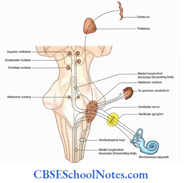
Afferent Connections of Vestibular Nuclei
- The vestibular nuclei receive afferents from vestibular receptors through the vestibular nerve, cerebellum, and vestibular nuclei of the opposite side.
- The vestibular pathway consists of first-order neurons (bipolar neurons), second-order neurons (vestibular nuclei), and third-order neurons (in the thalamus).
- The third-order neurons of this pathway project to the postcentral gyrus, the cortical area for vestibular sensation.
Efferent Connections of Vestibular Nuclei
The efferents from vestibular nuclei project to the cerebellum, brainstem (motor nuclei of cranial nerves), spinal cord, and cerebral cortex.
Vestibulospinal Tract
- The vestibulospinal tract originates from the cells of the lateral vestibular nucleus.
- The fibers of this tract are uncrossed and descend in the medulla dorsal to the inferior olivary nucleus and continue throughout the spinal cord in the ventral funiculus.
- The vestibulospinal fibers terminate in the anterior horn cells (motor neurons) that supply skeletal muscles.
- This tract is concerned with the maintenance of balance by regulating the tone of the muscles involved in posture.
Medial Longitudinal Fasciculus
- The axons of medial and inferior vestibular nuclei descend in the MLF (medial vestibulospinal tract) of both sides.
- These fibers travel through the floor ofthe fourth ventricle and medulla to terminate in the cervical part of the spinal cord.
- This tract influences the cervical motor neurons which move the head in such a way that equilibrium and fixation of gaze are maintained.
Medial Longitudinal Fasciculus Functions
- The vestibular nerve is concerned with conveying impulses associated with equilibrium The hair cells in the ampulla of the semicircular canal are sensors of kinetic balance (rotation of the head in any plane).
- The hair cells of the utricle are sensors of changes in gravitational forces, linear acceleration in the long axis of the body, and position of the head in space (i.e. static balance).
- The hair cells of the saccule are sensors of linear acceleration in the ventrodorsal axis of the body.
Stimulation of Labyrinth
- Vertigo is a sensation of movement in which the surrounding environment seems to revolve.
- It is a common symptom of the disease of the vestibular system. Probably, the cortical projections are responsible for the sense of vertigo.
Motion sickness: It is characterized by many symptoms such as nausea, headache, dizziness, and vomiting.
- This disease is caused due to motion during travel by road, sea, or air.
- Motion sickness occurs due to different messages received by the vestibular apparatus and eyes.
- For example, while traveling in a vehicle, the vestibular apparatus senses the motion but the eyes looking at the interior of the vehicle perceive it as still. These conflicting messages give the feeling of nausea.
Auditory And Vestibular Systems Summary
Hearing and equilibrium are two special somatic senses. The cochlear part of the membranous labyrinth is concerned with the reception of sound waves while the vestibular part of the labyrinth contains receptors for equilibrium.
Auditory system
- The cochlear nerve ends in dorsal and ventral cochlear nuclei.
- The axons of cochlear nuclei terminate on the superior olivary nucleus.
- The axons of the superior olivary nucleus form the lateral lemniscus.
- The lateral lemniscus ends on the neurons of the inferior colliculus whose fibers terminate on the medial geniculate body.
- The fibers of the medial geniculate body form auditory radiation which terminates on the auditory cortex.
Vestibular system
- The vestibular nuclei are present in the lower pons and upper medulla beneath the ‘vestibular area’ of the floor of the fourth ventricle.
- The vestibular part of the vestibulocochlear nerve ends in these nuclei.
- The afferent connectors of vestibular nuclei are from the vestibular nerve, from the cerebellum, and the opposite vestibular nuclei.
- The efferent connections of vestibular nuclei go to the cerebellum, brainstem, spinal cord, and cerebrum.
Auditory And Vestibular Systems Multiple Choice Questions
Question 1. Which of the following statements is false?
- Hearing and equilibrium are two special visceral senses
- The receptors for these two special senses are located in the membranous labyrinth
- The membranous labyrinth is located in the internal ear
- The cochlea is concerned with hearing
- The vestibular part of the membranous labyrinth has receptors for equilibrium
Answer: 1. Hearing and equilibrium are two special visceral senses
Question 2. Which of the following steps is false in the transmission of sound from the tympanic membrane to the cochlea?
- Vibration of the tympanic membrane
- Vibration of malleus, incus and stapes
- Vibration of the membrane covering the round window
- Vibration of perilymph of scala vestibule and scala tympani
- Vibration of the basilar membrane
Answer: 3. Vibration of perilymph of scala vestibule and scala tympani
Question 4. Which are the parts of a membranous labyrinth?
- Utricle
- Saccule
- Semicircular canals
- Cochlea
- All of the above
Answer: 1. Utricle
Question 5. Which of the following functional components are present in the cochlear nerve?
- General somatic efferent
- Special visceral efferent
- General somatic efferent
- Special somatic efferent
Answer: 3. General somatic efferent
Question 5. Which of the following ganglia/nuclei is not involved in the auditory pathway?
- Bipolar cells of the spiral ganglion
- Cochlear nuclei
- Inferior olivary nucleus
- Inferior colliculus
- Medial geniculate body
Answer: 3. Inferior colliculus
Question 6. Which of the following is not a part of the vestibular apparatus?
- Scala vestibule
- Utricle
- Saccule
- Semicircular ducts
Answer: 1. Scala vestibule
Question 7. The vestibular area consists ofthe following nuclei except
- Superior
- Inferior
- Medial
- Lateral
- Dorsal
Answer: 5. Dorsal